수면 장애 및 아토피피부염에 효과적인 7 가지 한약 처방의 항염증 활성 연구
Ⓒ The Society of Pathology in Korean Medicine, The Physiological Society of Korean Medicine
Abstract
This study was conducted to find a candidate prescription with anti-inflammatory efficacy of 7 herbal prescriptions known to be effective in atopic dermatitis and sleep disorders in Korean medicine. The anti-inflammatory of the 7 herbal prescriptions extracts were evaluated by ELISA assay and Western blot assay. 7 herbal prescriptions, Seongyutang(SU), Danseontoetang(DST), Sotosajahwan(ST), Jisiljagyagsan(JJ), Seokchangpo(SCP), Wiryeongseon(WLS), Gogojohwan(GGJ) 30 % EtoH extract was used for in vitro experiments. In TNF-α + IFN-γ(TI) stimulated HaCaT cells, all 7 herbal prescriptions reduced TARC and MDC production, and JJ strongly reduced TARC and MDC production at 100 ㎍/ml concentration. SCP strongly reduced MDC production at 10 ㎍/ml and 100 ㎍/ml concentration. In addition, in substance P(SP)/CRH stimulated HMC-1 cells, JJ strongly inhibited VEGF production at both 10 and 100 ㎍/ml concentrations. In LPS/CRH stimulated Raw264.7 cells, all 7 herbal prescriptions significantly inhibited TNF-α. PMA + Ionomycin(PI)/CRH stimulated EL4 cells, SU significantly reduced IL-4 production at both concentrations of 10 and 100 ㎍/ml. WLS significantly reduced IL-17 production at both concentrations of 10 and 100㎍/ml. This suggests that 7 herbal prescriptions have anti-inflammatory effects by reducing inflammatory cytokines in keratinocytes, mast cells and macrophages caused by inflammation. Therefore, it is expected that 7 herbal prescriptions that are effective for sleep disorders and atopic dermatitis can be used as therapeutic materials for sleep disorders and diseases associated with atopic dermatitis.
Keywords:
Atopic dermatitis, Sleep disturbance, Herbal prescriptions, Comorbidity, Stress서 론
아토피피부염(Atopic dermatitis, AD)은 홍반, 부종, 태선화 등의 피부 증상과 소양감을 호소하는 만성 염증성 피부 질환이다.1) 아토피피부염 환자는 질병 이환 기간 동안 수면장애를 동반하는 경우가 많으며, 이로 인한 피로감, 기분 장애, 업무 수행 능력 저하, 삶의 질 저하 등을 경험하는 것으로 보고되었다.2,3)
6세 이하 300 명의 아토피피부염 소아 환자를 대상으로 한 연구결과, 60% 이상에서 수면장애를 경험하고 있어 아토피피부염 환자의 관리를 위해 수면장애를 개선하는 치료 전략이 필요함을 시사하고 있다.4)
아토피 피부염은 한의학에서 奶癬, 嬰兒濕疹, 浸淫瘡, 胎癬瘡, 血風瘡 등의 범주에 속한다.5) 胎火濕熱이 蘊積하거나 風熱之邪가 침입하여 아토피피부염이 유발되는 것으로 보고 있으며, 風熱, 濕熱, 血熱, 虛熱 등으로 변증하여 치료한다.6) 서양의학에서 아토피피부염 치료를 위해 보습제, 스테로이드, 면역억제제 사용 등을 사용하고 있으나, 사용 중 불규칙한 수면시간, 입면 후 잠자리에 드는 시간이 오래 걸림, 잠자리 시 자주 깸 등을 호소하는 사례가 보고되고 있다.7,8) 최근 消風淸營湯加味方, 升麻黃連湯 등의 처방을 활용하여 아토피피부염 환자에 동반된 수면장애 증상을 개선한 사례가 보고되었다.9) 또한, 불면증, 야제증 등에 사용되는 抑肝散을 활용한 실험 연구에서 TNF-α, IL-4등의 염증성 cytokine 발현을 억제하여 아토피 피부염 증상을 개선한 사례가 보고되었다.10)
따라서 본 연구에서는 한의학 고전에서 아토피 피부염 증상 및 수면장애를 동시에 치료할 수 있을 것으로 예상되는 한약 처방을 탐색한 후, 피부각질형성세포, 비만세포, 대식세포에서 항염증효능을 관찰하였다. 이에 대한 유의한 결과를 얻었기에 보고하는 바이다.
재료 및 방법
1. 재료 및 시약
본 실험에 사용한 Human mast cell HMC-1 cell과 Mouse T cell EL4 cell, Mouse macrophage Raw 264.7 cell은 한국세포주은행(Seoul, South Korea)에서 구입하였다. 또한 Iscove’s modified Dulbecco’s 배지(IMDM)은 Merck Millipore(Darmstadt, Germany)와 High glucose Dulbecco’s modified Eagle’s 배지(DMEM)은 Welgene Inc.(Gyeongsangbuk, Korea)을 사용하였으며, 10% Fetal bovine serum(FBS) 및 1% Penicilin, Streptomycin(PS)은 Invitrogen Inc.(Carlsbad, CA, USA)에서 구입하여 사용하였다. Human vascular endothelial growth factor(VEGF) 및 Mouse tumor necrosis factor(TNF)-α, interleukin(IL)-4, interleukin(IL)-17 ELISA Kit는 Koma Biotech Inc.(Seoul, Korea)에서 구입하였고, human thymus and activation-regulated chemokine(TARC/CCL17)과 macrophage-derived chemokine(MDC/CCL22) ELISA Kit는 R&D Systems(Minneapolis, MN, USA)에서 구입하였다.
2. 7가지 한약 처방 선택
發斑, 腫脹, 乾騷, 濕瘡, 癬, 痒 등의 아토피피부염 증상 관련 용어와 不眠, 不得臥, 多夢 등의 수면장애 증상 관련 용어 목록 작성 후, 고문헌에서 주치 병증, 방제명, 처방 구성의 정보를 가지는 데이터베이스를 구축하였다. 동의보감, 의학입문, 경악전서, 광제비급, 동의수세보원, 제중신편, 방약합편, 향약집성방의 원문텍스트를 이용하여 검색하였으며, 이 중 임상활용도가 높은 7가지 처방을 선택하였다. 이를 토대로 아토피피부염 및 수면장애 증상 용어 목록의 내용이 방제의 주치 병증에 포함된 경우를 검색하여 공통적으로 사용할 수 있는 방제 목록을 추출, 분석하였다. 추출된 방제 목록을 이용하여 본초 구성, 주치 병증 정보를 함께 출력하였다. 도출한 7종의 처방에 사용된 한약재는 강동경희대학교 한방병원에서 구입하였다(Table 1).
3. 약재 구입 및 추출물 제조
성유탕(SU), 단선퇴탕(DST), 소토사자환(ST), 지실작약산(JJ), 석창포(SCP), 위령선(WLS), 고고조환(GGJ)을 각각 30% EtOH로 80℃ 조건에서 3시간 동안 전탕 후 0.2mm 필터(Whatman International, Maidstone, UK)로 여과하여 불용성 물질을 제거하였다. 여과된 추출액은 Rotary vacuum evaporator을 사용해서 1시간 동안 농축한 후, 이를 -80℃ 동결건조기(FD8508S, Busan, Korea)에서 3일 동안 건조시켰다. 모든 시료는 추가 연구를 위해 -20℃에 보관하였다.
4. 세포배양
HMC-1 cell은 1% PS와 10% FBS, 1.2mM 1-thiolglycerol이 포함된 IMDM 배지를 사용하여 배양하였으며, EL4 cell, HaCaT cell, Raw 264.7 cell은 1% PS와 10% FBS가 포함된 DMEM 배지를 사용하여 37℃, 5% CO2 조건에서 배양하였다. 실험에 사용된 모든 세포는 80-90% confluence로 자랐을 때 계대 배양하였다.
5. Cell viability assay
실험에 사용된 모든 세포는 언급된 배지에 5 x 10⁴ cells/ml의 농도로 섞은 후, 96 well plate에 100 ㎕씩 분주하고 37℃, 5% CO2 조건에서 24시간 동안 배양하였다. 이후 10, 100, 500, 1000 ㎍/ml 농도로 추출물을 처리하고 24시간 동안 배양한 뒤, 2, 3-Bis (2-methoxy-4-nitro-5-sulfophenyl)-2H-tetrazolium-5-carboxanilide(XTT) 시약을 각 well 당 50 ㎕씩 첨가하여 4시간 동안 CO2 incubator에서 반응시켰다. 이후 Microplate reader(Tecan, Männedorf, Switzerland)를 사용하여 450 nm에서 흡광도를 측정하였다. 세포생존율은 정상 대조군의 백분율로 비교하였다.
6. Enzyme-linked Immunosorbent Assay(ELISA)
HMC-1 cell을 12 well plate에 각 well 당 5 x 10⁵ cells/ml의 농도로 분주한 뒤, 1시간 동안 37℃, 5% CO2조건에서 배양하였다. 추출물 10, 100 ㎍/ml과 Dexamethasone 10 ㎛을 1시간 전 처리하고 Substance P(SP) 10 ㎛, Corticotropin-releasing hormone(CRH) 1 nM을 각각 48시간, 24시간 동안 처리하여 배양하였다. 이후 배지를 1,500 rpm, 10분 동안 원심 분리하여 얻은 상층액을 사용하여 VEGF 생성 함량을 측정하였다.
EL4 cell을 12 well plate에 각 well 당 5 x 10⁵ cells/ml의 농도로 분주한 뒤, 1시간 동안 37 ℃, 5% CO2조건에서 배양하였다. 추출물 10, 100 ㎍/ml과 Dexamethasone 10 ㎛을 1시간 전처리한 다음, phorbol myristate acetate(PMA) 10 ng/ml + Ionomycin 100 ng/ml(PI)을 처리하고 1시간 후에 CRH 10 nM을 처리하여 24시간 동안 배양하였다.
HaCaT cell을 6 well plate에 각 well 당 5x10⁵ cells/ml의 농도로 분주한 뒤, 24시간 동안 37℃, 5% CO2조건에서 배양하였다. FBS가 포함되지 않은 배양액에서 24시간 동안 재 배양한 후에 추출물 10, 100 ㎍/ml과 Dexamethasone 10 ㎛을 1시간 전처리 한 다음, TNF-α, interferon-gamma(INF-γ) 10 ng/ml을 각각 처리 후 24시간 배양하였다.
Raw 264.7 cell을 12 well plate에 각 well 당 2.5x10⁵ cells/ml의 농도로 분주한 뒤, 24시간 동안 37℃, 5% CO2조건에서 배양하였다. FBS가 포함되지 않은 배양액에서 24시간 동안 재 배양한 후에 추출물 10, 100 ㎍/ml과 Dexamethasone 10 ㎛을 1시간 전처리 한 다음, lipopolysaccharide(LPS) 1 ㎍/ml을 처리하고 1시간 후 CRH 1 nM을 처리하여 24시간 배양하였다.
결 과
1. 한약 처방 추출물이 HaCaT세포의 TARC/CCL17, MDC/CCL22 생성에 미치는 영향
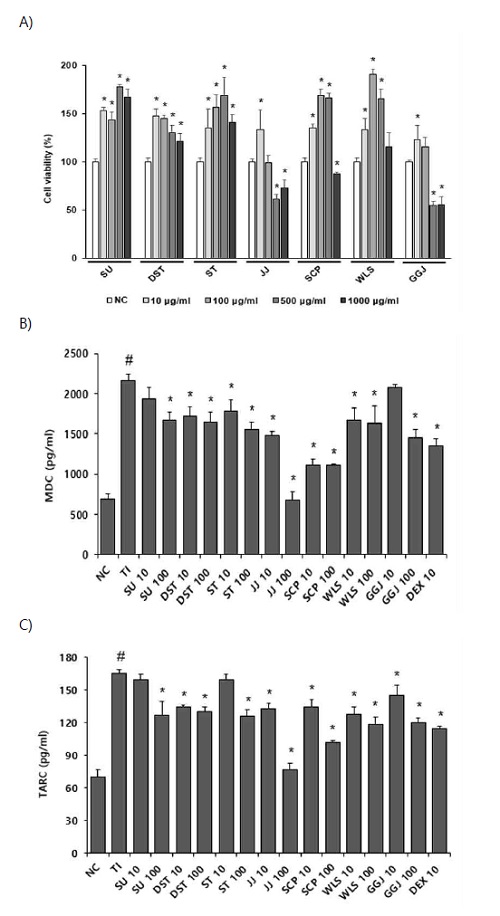
HaCaT 세포에 SU, DST, ST, JJ, SCP, WLS, GGJ 처방 추출물을 10, 100, 500, 1000 ㎍/ml의 농도로 처리하고 24시간 후에 XTT assay를 통해 세포 생존율을 확인하였다. SU, DST, ST, JJ, SCP, WLS, GGJ 처방 추출물에서 100 ㎍/ml의 농도까지는 90% 이상의 세포 생존율을 보였으며 JJ, GGJ 처방 추출물은 500 ㎍/ml 이상의 농도를 처리하였을 때 80% 이하로 세포 생존율이 감소하였다. 따라서 모든 처방 추출물에서 독성이 나타나지 않은 10, 100 ㎍/ml의 농도에서 실험을 진행하였다(Fig. 1A). 항 아토피 효능 평가는 HaCaT 세포에 TI 처리를 통하여 염증 반응을 일으키고 SU, DST, ST, JJ, SCP, WLS, GGJ 처방 추출물을 10, 100㎍/ml 농도로 처리하여 배양 후 TARC/CCL17, MDC/CCL22의 생성 억제를 측정하였다. 세포 상층액에서 MDC의 발현을 측정한 결과 TI 처리군과 비교하여 SU(100 ㎍/ml), DST(10, 100 ㎍/ml), ST(10, 100 ㎍/ml), JJ(10, 100 ㎍/ml), SCP(10, 100 ㎍/ml), WLS(10, 100 ㎍/ml), GGJ(100 ㎍/ml)를 처리했을 때 MDC의 발현이 유의미한 감소를 보였다(Fig. 1B). 또한, SU(100 ㎍/ml), DST(10, 100 ㎍/ml), ST(100 ㎍/ml), JJ(10, 100 ㎍/ml), SCP(10, 100 ㎍/ml), WLS(10, 100 ㎍/ml), GGJ(10, 100 ㎍/ml)를 처리했을 때 TARC 발현이 유의미한 감소를 보였다(Fig. 1C).

Effect of prescription on the MDC and TARC expressions in TI-stimulated HaCaT cells. (A) HaCaT cells were treated with 10, 100, 500, 1000 ㎍/ml of prescription extracts for 24h. Cell viability was assessed using XTT assay. (B,C) HaCaT cells were 1h pre-treated with prescription extracts (10, 100 ㎍/ml) and DEX 10㎛, then stimulated with TNF-α, INF-γ 10 ng/ml each for 24h. Levels of MDC and TARC in culture supernatants were measured by ELISA assay. (#p < 0.05 vs. NC *p <0.05 vs TI treated group)
2. 한약 처방 추출물이 HMC-1세포의 VEGF 생성에 미치는 영향
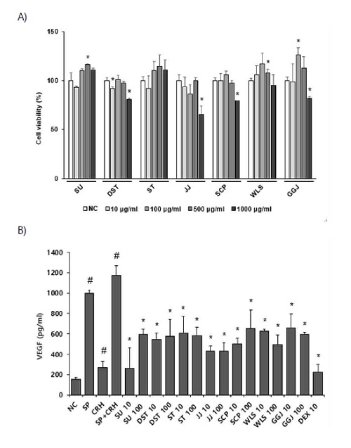
HMC-1 세포에서 처방 추출물의 세포 독성 효과를 알아보기 위하여 HaCaT 세포와 동일한 방법으로 세포의 생존율을 확인하였다. SU, DST, ST, JJ, SCP, WLS, GGJ처방 추출물에서10, 100, 500 ㎍/ml의 농도까지는 90% 이상의 세포 생존율을 보였으며DST, JJ, SCP, GGJ 처방 추출물은 1000 ㎍/ml의 농도를 처리하였을 때 80% 이하로 세포 생존율이 감소하였다. HMC-1 세포에 SP + CRH 처리를 통하여 염증 반응을 일으키고 SU, DST, ST, JJ, SCP, WLS, GGJ 처방 추출물을 10, 100 ㎍/ml 농도로 처리하여 배양 후 VEGF 생성 억제를 측정하였다. 세포 상층액에서 VEGF의 발현을 측정한 결과 SP + CRH 처리군과 비교하여 SU(10, 100 ㎍/ml), DST(10, 100 ㎍/ml), ST(10, 100 ㎍/ml), JJ(10, 100 ㎍/ml), SCP(10, 100 ㎍/ml), WLS(10, 100 ㎍/ml), GGJ(10, 100 ㎍/ml)를 처리했을 때 VEGF의 발현이 유의미한 감소를 보였다(Fig. 2B).

Effect of prescription on the VEGF expressions in SP and CRH-stimulated HMC-1 cells. (A) HMC-1 cells were treated with 10, 100, 500, 1000 ㎍/ml of prescription extracts for 24h. Cell viability was assessed using XTT assay. (B) HMC-1 cells were 1h pre-treated with prescription extracts 10, 100 ㎍/ml and DEX 10㎛ then stimulated with SP 10㎛ for 48h. next day, stimulated with CRH 1nM for 24h. Levels of VEGF in culture supernatants were obtained by centrifuging the medium at 1,500 rpm for 10 minutes is used measured by ELISA assay. (#p < 0.05 vs. NC *p <0.05 vs SP and CRH treated group).
3. 한약 처방 추출물이 Raw 264.7 세포의 TNF-α 생성에 미치는 영향
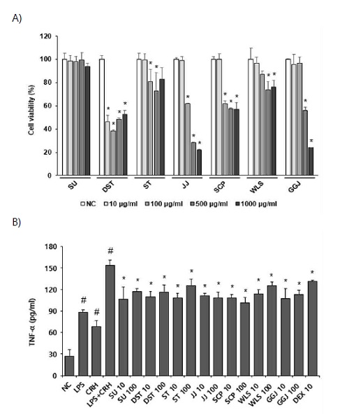
Raw264.7 세포에 SU, DST, ST, JJ, SCP, WLS, GGJ 처방 추출물을 10, 100, 500, 1000 ㎍/ml의 농도로 처리하고 24시간 후에 XTT assay를 통해 세포 생존율을 확인하였다. SU, ST, JJ, SCP, WLS, GGJ 처방 추출물에서 10 ㎍/ml의 농도까지는 90% 이상의 세포 생존율을 보였다. 한편 각각 처방 추출물 JJ, SCP는 100 ㎍/ml 이상의 농도에서 80% 이하의 세포 생존율과 ST, WLS, GGJ는 500 ㎍/ml 이상의 농도에서 80% 이하의 생존율을 보였다. DST 처방 추출물은 10 ㎍/ml 이상의 농도를 처리하였을 때 50% 이하로 세포 생존율이 감소하였다(Fig. 3A). DST는 10 ㎍/ml의 농도에서도 독성이 나타났지만 모든 세포의 조건을 동일하게 하기 위해 실험은 10, 100 ㎍/ml의 농도에서 진행하였다. Raw264.7 세포에 LPS+CRH 처리를 통하여 염증 반응을 일으키고 SU, DST, ST, JJ, SCP, WLS, GGJ 처방 추출물을(10, 100 ㎍/ml) 농도로 처리하여 배양 후 TNF-α 생성 억제를 측정하였다. 세포 상층액에서 TNF-α의 발현을 측정한 결과 LPS+CRH 처리군과 비교하였을 때 SU(10, 100 ㎍/ml), DST(10, 100 ㎍/ml), ST(10, 100 ㎍/ml), JJ(10, 100 ㎍/ml), SCP(10, 100 ㎍/ml), WLS(10, 100 ㎍/ml), GGJ(10, 100 ㎍/ml)를 처리했을 때 TNF-α의 발현이 유의미한 감소를 보였다.

Effect of prescription on the TNF-α expressions in LPS and CRH-stimulated Raw264.7 cells. (A) Raw264.7 cells were treated with 10, 100, 500, 1000 ㎍/ml of prescription extracts for 24h. Cell viability was assessed using XTT assay. (B) Raw264.7 cells were 1h pre-treated with prescription extracts 10, 100 ㎍//ml and DEX 10㎛ then stimulated with LPS 1ng/ml for 24h. after 1h, stimulated with CRH 1nM for 24h. Levels of MDC and TARC in culture supernatants were measured by ELISA assay. (#p < 0.05 vs. NC *p <0.05 vs LPS and CRH treated group).
4. 한약 처방 추출물이 EL4 세포의 IL-4, IL-17 생성에 미치는 영향
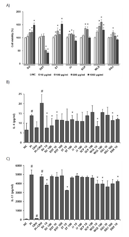
EL4 세포에서 처방 추출물의 세포 독성 효과를 알아보기 위하여 HaCaT 세포와 동일한 방법으로 세포의 생존율을 확인하였다. SU, DST, ST, JJ, SCP, WLS, GGJ 처방 추출물에서 100 ㎍/ml의 농도까지는 90% 이상의 세포 생존율을 보였다. 한편 DST 처방 추출물은 500 ㎍/ml 이상의 농도를 처리하였을 때 50% 이하로 세포 생존율이 감소하였다. 따라서 모든 처방 추출물에서 독성이 나타나지 않은 10, 100 ㎍/ml의 농도에서 실험을 진행하였다(Fig. 4A). EL4 세포에 PI + CRH 처리를 통하여 염증 반응을 일으키고 SU, DST, ST, JJ, SCP, WLS, GGJ 처방 추출물을 10, 100 ㎍/ml 농도로 처리하여 배양 후 IL-4와 IL-17 생성 억제를 측정하였다. 세포 상층액에서 IL-4 발현을 측정한 결과 PI + CRH 처리군과 비교하여 SU(10, 100 ㎍/ml), DST(10 ㎍/ml), ST(100 ㎍/ml), WLS(10 ㎍/ml), GGJ(100 ㎍/ml)을 처리했을 때 IL-4의 발현이 유의미한 감소를 보였다(Fig. 4B). 한편 IL-17 발현을 측정한 결과 PI + CRH 처리군과 비교하여 SU(10 ㎍/ml), ST(10 ㎍/ml), WLS(10, 100 ㎍/ml)을 처리했을 때 IL-17의 발현이 유의미한 감소를 보였다. 반면 SU(100 ㎍/ml), DST(10, 100 ㎍/ml), ST(100 ㎍/ml), JJ(10, 100 ㎍/ml), SCP(10, 100 ㎍/ml), GGJ(10, 100 ㎍/ml)에서는 IL-17의 생성에 관하여 유의미한 억제 효과를 나타내지 않았다(Fig. 4C).

Effect of prescription on the IL-4 and IL-17 expressions in PI and CRH-stimulated EL4 cells. (A) EL4 cells were treated with 10, 100, 500, 1000 ㎍/ml of prescription extracts for 24h. Cell viability was assessed using XTT assay. (B,C) EL4 cells were 1h pre-treated with prescription extracts 10, 100 ㎍/ml and DEX 10㎛ then stimulated with PMA 10 ng/ml, Ionomycin 100 ng/ml each for 24h. after 1h, stimulated with CRH 10nM for 24h. Levels of IL-4 and IL-17 in culture supernatants were measured by ELISA assay. (#p < 0.05 vs. NC *p <0.05 vs PI and CRH treated group).
고 찰
아토피피부염은 만성 재발성 피부질환으로 환자의 외적인 변화를 초래함으로써 자존감이 낮아지고 불안, 우울과 같은 정신적 문제를 초래할 수 있다.11) AD 환자에서 소양감으로 인한 수면장애는 매우 흔하게 발생하고 정상인에 비해 수면 효율이 약 4배 낮으며, non-rapid eye movement(non-REM) 수면 수치가 낮게 나타난다.12) 이와 같이 아토피피부염 환자에서 수면장애는 피로감이나 삶의 질을 저하시키고 정신적 스트레스 등에 영향을 주어 사회적 문제로도 이어질 수 있다.13) 정신적 스트레스는 불면증, 우울증과 같은 정신 질환의 발생을 촉진 할뿐만 아니라 병원체 감염 질환에 대한 감수성을 크게 증가시킨다.14)
수면장애는 Hypothalamic - Pituitary - Adrenal(HPA) 축의 조절 이상을 유발해 신경 전달 물질인 세로토닌을 감소시키고 CRH및 Cortisol의 수준을 증가시킨다.15) CRH는 HPA 축의 활성화와 glucocorticoids 및 catecholamines의 방출을 통해 신경 내분비 -면역 상호 작용에 영향을 미친다.16) 특히, 스트레스호르몬인 CRH는 비만세포에 영향을 미쳐 히스타민의 탈과립 및 염증 유발물질인 히스타민의 방출을 유도한다.17)
역학 및 보고된 연구에 따르면 스트레스 호르몬은 Th2를 자극하여 알레르기, 염증성 피부질환에 영향을 미친다고 보고되었다.18) 그 중에서 MDC와 TARC는 대표적인 Th2 케모카인으로 CC chemokine receptor 4(CCR4) 에 작용하여 표적 장기로 Th2 세포의 이동과 침윤을 유도하는 것으로 알려져 있다.19) 케모카인에 의해 표적 장기로 이주한 Th2 세포는 B 세포를 자극하여 IL-4, IL-5, IL-13과 같은 사이토카인을 분비하여 알레르기 염증반응을 강화하고 MDC와 TARC의 생성을 자극하여 지속적으로 Th2 세포의 이동을 자극하여 만성적인 염증 상태가 지속되도록 한다.20) 따라서 본 연구에서 피부각질형성세포인 HaCaT 세포에서 TI를 처리하여 염증을 유도하고 아토피피부염 유사 환경을 조성하였으며, 대조군에 비해 TI처리군에서 MDC와 TARC의 생성량이 유의미하게 증가하였다. 이는 처방 추출물을 처리하였을 때 DST, ST, JJ, SCP, WLS에서 MDC의 생성량이 유의미하게 감소하였고 특히, SCP는 처리한 농도 모두에서 MDC의 생성량이 크게 감소하였다. 또한 DST, JJ, SCP, WLS, GGJ에서 TARC의 생성량이 유의미하게 감소하였으며 JJ, SCP 100 ㎍/ml의 농도에서TARC 생성량이 크게 감소하였다. 따라서 본 실험을 통해 처방 추출물의 항염증효능을 확인하였으며 이에 아토피피부염에 효과가 있다고 보고된 처방 추출물은HaCaT 세포에서 TI에 의해 유발된 염증 반응에 MDC와 TARC 생성량 억제를 통하여 염증세포의 침윤을 조절함으로써 아토피피부염 증상 완화에 기여할 수 있을 것으로 사료된다.
비만 세포는 주로 결체조직과 점막에 존재하면서 알레르기와 염증 반응에 관여하고 IgE(FcεRI)에 대한 고친화성 수용체의 교차 결합에 반응하여 과립 내의 사이토카인을 분비하여 알레르기 염증의 초기 반응과 후기 반응을 일으키고 만성적인 염증 반응에 주요한 역할을 한다.21) 또한 비만 세포는 세포질 과립 내에 VEGF를 포함하고 있으며, 이 성장 인자를 자발적으로 그리고 활성화 후에 분비 할 수 있다.22) 스트레스 호르몬인 CRH는 비만 세포의 CRH 수용체에 결합하여 VEGF 분비량을 증가하고, 이는 혈관투과도를 증가하여 국소 염증반응을 강화한다.23) 따라서 본 연구에서 인간비만세포주인 HMC-1 세포에서 스트레스 호르몬인 CRH와 SP를 처리하여 염증을 유도하였다. 대조군에 비해 SP+CRH처리군에서 VEGF 생성량이 증가하였으며 이는 처방 추출물을 처리하였을 때 유의미하게 감소하였다. 특히 SU 10 ㎍/ml의 농도에서 VEGF 생성량이 가장 크게 감소하였다.
대식세포는 염증반응에서 방어에 중추역할을 하고 LPS를 인지한 염증반응은 전사 인자인 NF-κB 조절에 관여한다.24) 이를 통해서 염증성 사이토카인의 생산과 분비를 자극하고 염증 촉진 매개물질을 유도한다. 자극을 받은 대식세포로 인해 NF-κB가 활성화되면 TNF-α, IL-6 등을 증가시켜 염증반응을 유도한다.25) 따라서 본 연구에서 대식세포인 Raw264.7 세포에서 스트레스 호르몬인 CRH와 LPS를 처리하여 염증을 유도하였다. 대조군에 비해 LPS +CRH 처리군에서 TNF-α 생성량이 증가하였으며 이는 처방 추출물을 처리하였을 때 유의미하게 감소하였다. 하지만 DST의 경우 모든 농도에서 세포 생존율이 50% 이하로 감소한 것으로 보아 10, 100 ㎍/ml의 농도에서 TNF-α의 발현의 감소는 세포 독성으로 인한 결과로 사료된다. 또한 JJ, SCP는 100 ㎍/ml 농도에서 세포 생존율이 80% 이하인 것으로 보아 100 ㎍/ml의 농도에서 TNF-α의 발현의 감소는 세포 독성으로 인한 결과로 사료된다.
항원을 제공받은 T 세포는 Th2 세포로 분화하며 IL-4, IL-13, IL-17 등과 같은 사이토카인을 분비하여 염증을 유발하고, 면역 세포를 자극해서 IgE 및 염증 매개 물질들이 분비됨으로써 과민반응이 유발된다.26,27) 따라서 본 연구에서 마우스 T 세포인 EL4 세포에서 스트레스 호르몬인 CRH와 PMA/Ionomycin(PI)를 처리하여 염증을 유도하였다. 대조군에 비해 PI +CRH 처리군에서 IL-4, IL-17의 생성량이 유의미하게 증가하였다. 이는 처방 추출물 SU, DST, ST, WLS, GGJ을 처리했을 때 IL-4의 발현이 유의미한 감소하였고, SU, ST, WLS을 처리했을 때 IL-17의 발현이 유의미한 감소를 보였다. 따라서 PI +CRH로 유도된 T세포의 in vitro 모델에서 처방 추출물 SU(10, 100 ㎍/ml), DST(10 ㎍/ml), ST(100 ㎍/ml), WLS(10 ㎍/ml), GGJ(10 ㎍/ml)의 농도에서 염증성 사이토카인 IL-4와 SU(10 ㎍/ml), SU(10 ㎍/ml), WLS(10, 100 ㎍/ml)의 농도에서 IL-17 발현을 감소시키는 것으로 보아, 아토피피부염과 연관된 수면장애 개선 효과에 기여할 수 있을 것으로 사료된다. 7가지 처방 중 18종의 약물(산약, 감초, 고삼, 당귀, 토사자, 백복령, 생지황, 연자육, 석창포, 선퇴, 숙지황, 위령선, 인삼, 작약, 조협, 지실, 천궁, 황기)이 사용되었으며, 이중 補益藥에 해당하는 약물이 8종 (산약, 감초, 당귀, 토사자, 숙지황, 인삼, 작약, 황기)으로 가장 많았다. 일반적으로 한의학에서는 아토피피부염의 원인을 風熱, 血熱 등의 實한 상황으로 본다. 그와 반대로 補益藥이 다빈도 처방이라는 점에서, 수면장애가 동반되는 AD 상황에서는 그 원인을 해석할 때 虛證 관련으로 보고, 補益하는 여러 방법으로 치료할 수 있을 것이다.28) 수면장애가 동반되는 아토피피부염 치료 시 後天之本으로서 精血 생성의 원천이 되는 脾胃의 기능을 다룸으로써 증상을 개선시킬 수 있을 것이라 예상된다.29) 본 연구에서 사용한 한약처방 7종의 효능에 대해 in vitro 수준에서의 실험만 이루어졌다. 따라서 동물 모델을 활용한 효력 시험, 약동력학 및 안전성 연구 등을 포함한 추가적 연구가 필요하다. 또한 IL-17 발현을 알아본 실험의 결과 중 일부 처방에서 유의미한 결과가 나타나지 않았으므로 이에 대한 추가적인 연구가 진행되기를 희망한다.
결 론
한의학 고전에서 아토피피부염과 수면장애에 효과가 있다고 알려진 7가지 한약 처방을 탐색한 후 항염증 효과를 측정한 결과 HaCaT 세포에서 MDC, TARC 생성을 억제하였고 HMC-1 세포와 Raw264.7 세포에서 각각 VEGF와 TNF-α의 분비를 억제하였다. 또한 EL4 세포에서 7가지 한약처방 중 SU, ST, WLS의 일부 농도에서 IL-4와 IL-17의 생성을 억제하여 스트레스 상황을 동반한 아토피피부염 유사 in vitro 모델에서 염증을 개선하는데 유의미한 효과를 보였다. 결과적으로 한약 처방 SU가 모든 세포에서 염증성 사이토카인의 발현을 감소시키는 것으로 보아 수면장애 및 아토피피부염 관련 질환의 치료제로 가장 우수한 처방이라 생각된다.
Acknowledgments
본 연구는 2020년도 과학기술정보통신부의 재원으로 한국연구재단 (NO. NRF-2019R1F1A1059856)과 동국대학교의 지원(2020)을 받아 수행된 연구결과입니다.
References
- Lee HY, Lee JR, Roh JY. Epidemiological Features of Preschool Childhood Atopic Dermatitis in Incheon. Korean Journal of Dermatology. 2009;47(2):164-71.
-
Bender BG, Ballard R, Canono B, Murphy JR, Leung DY. Disease severity, scratching, and sleep quality in patients with atopic dermatitis. J. Am. Acad. Dermatol. 2008;58(3):415-20.
[https://doi.org/10.1016/j.jaad.2007.10.010]

-
Bender BG, Leung SB, Leung DY. Actigraphy assessment of sleep disturbance in patients with atopic dermatitis: an objective life quality measure. J. Allergy Clin. Immunol. 2003;111(3):598-602.
[https://doi.org/10.1067/mai.2003.174]

-
Chamlin SL, Mattson CL, Frieden IJ, et al. The price of pruritus: sleep disturbance and cosleeping in atopic dermatitis. Arch. Pediatr. Adolesc. Med. 2005;159(8):745-50.
[https://doi.org/10.1001/archpedi.159.8.745]

- Yun H-J, Ko W-S. Clinical Study of Atopic Dermatitis ; the Classification of Oriental Medical Clinical type and Treatment. The Journal of Korean Medicine. 2001;22(2):10-21.
- Hye JJ, Moon LJ, Yeon LS. A Clinical Study of Atopic Dermatitis for Children. Journal of Korean Oriental Pediatrics. 2005;19(2):69-84.
-
Cho J-M, Park S-J, Lee H-T, Han S-R. Five Cases of the Patients with the Nummular Eczema Treated with Sihochunggan-tang gagambang and Sunbangpaedok-tang gagambang. Journal of Korean Med Ophthalmol Otolaryngol Dermatol. 2016;29(3):274-87.
[https://doi.org/10.6114/jkood.2016.29.3.274]

-
Su-Ryun H, Gun P, Myeong-Hwa H, et al. Retrospective Study about the Effectiveness of a long-term Korean Medicine Treatment on 511 Atopic Dermatitis Patients. The Journal of Korean Medicine Ophthalmology and Otolaryngology and Dermatology 2013;26(3):65-76.
[https://doi.org/10.6114/jkood.2013.26.3.065]

-
Song M-H, Lee J-H, Choi C-M. A Case Report of Atopic Eruption of Pregnancy by Traditional Korean Medicine. Journal of Korean Obstet Gynecol. 2016;29(3):91-9.
[https://doi.org/10.15204/jkobgy.2016.29.3.091]

-
Noh HM, Park SG, Jo EH, et al. Study about the Comparison of Korean-Western Medicine on Atopic Dermatitis and Psychological Factors. Journal of Physiology & Pathology in Korean Medicine. 2018;32(2):113-25.
[https://doi.org/10.15188/kjopp.2018.04.32.2.113]

- Lee HJ, Park CO, Lee JH, Lee KH. Life Quality Assessment among Adult Patients with Atopic Dermatitis. Korean Journal of Dermatology 2007;45(2):159-64.
-
Chang Y-S, Chiang B-L. Sleep disorders and atopic dermatitis: A 2-way street? J. Allergy Clin. Immunol. 2018;142(4):1033-40.
[https://doi.org/10.1016/j.jaci.2018.08.005]

- Ryu J-Y, Kam E-Y, Kang E-J, et al. Effects of Gammakdaejo-tang(GMD) on DNCB induced Atopic Dermatitis in Mice. The Journal of Korean Medicine Ophthalmology and Otolaryngology and Dermatology. 2020;33(2):83-99.
-
Pan M-H, Zhu S-r, Duan W, et al. "Shanghuo" increases disease susceptibility: Modern significance of an old TCM theory. J. Ethnopharmacol. 2019:112491.
[https://doi.org/10.1016/j.jep.2019.112491]

-
Cho S, Lee S. A study of longitudinal changes and other relevant factors of adolescents’ sleep duration. 2020;31:5~32.
[https://doi.org/10.14816/sky.2020.31.1.5]

-
Chrousos GP. The hypothalamic-pituitary-adrenal axis and immune-mediated inflammation. N Engl J Med. 1995;332.
[https://doi.org/10.1056/NEJM199505183322008]

-
Ito N, Sugawara K, Bodó E, et al. Corticotropin-releasing hormone stimulates the in situ generation of mast cells from precursors in the human hair follicle mesenchyme. J Invest Dermatol. 2010;130(4):995-1004.
[https://doi.org/10.1038/jid.2009.387]

- Ramírez F, Fowell DJ, Puklavec M, Simmonds S, Mason D. Glucocorticoids promote a TH2 cytokine response by CD4+ T cells in vitro. The Journal of Immunology. 1996;156(7):2406-12.
-
Hong S-J, Lee W-J, Jo E-H, Ahn S-H. The Effect of Lactobacillus Mixture Culture Fluid Extracts on Atopic Dermatitis Chemokine Expression of in HaCaT Cells. Korean Journal of Acupuncture. 2017;34(2):82-7.
[https://doi.org/10.14406/acu.2017.008]

-
Hartl D, Lee CG, Da Silva CA, Chupp GL, Elias JA. Novel biomarkers in asthma: chemokines and chitinase-like proteins. Curr. Opin. Allergy Clin. Immunol. 2009;9(1):60-6.
[https://doi.org/10.1097/ACI.0b013e32831f8ee0]

- Son JY, Bae HC, Renchinkhand G, Nam MS, Kim W-s. Effect of lactoferrin hydrolysates on inflammatory cytokine modulation in HEK-293, RBL-2H3, and HMC-1 cells. Korean Journal of Agricultural Science. 2020;47(1):83-93.
-
McHale C, Mohammed Z, Gomez G. Human Skin-Derived Mast Cells Spontaneously Secrete Several Angiogenesis-Related Factors. Front. Immunol. 2019;10(1445).
[https://doi.org/10.3389/fimmu.2019.01445]

-
Cao J, Papadopoulou N, Kempuraj D, et al. Human mast cells express corticotropin-releasing hormone (CRH) receptors and CRH leads to selective secretion of vascular endothelial growth factor. The Journal of Immunology. 2005;174(12):7665-75.
[https://doi.org/10.4049/jimmunol.174.12.7665]

- Kim S-J, kim T-J, Kim E-H, Kim Y-M. Anti-inflammatory and Anti-oxidant Studies of Osung-tang Extracts in LPS-Induced RAW 264.7 Cells. The Journal of Korean Medicine Ophthalmology and Otolaryngology and Dermatology. 2020;33(1):1-11.
- Park HJ, Bae GS, Kim DY, et al. Inhibitory Effect of Extract from Ostericum koreanum on LPS-induced Proinflammatory Cytokines Production in RAW264.7 Cells. Kor. J. Herbology. 2008;23(3):127-34.
-
Kim B-KL, Jong-Soon Kil, Ki-Jung. Effects of Imperatae Rhizoma Extract on T helper 2 cell differentiation. The Korea Journal of Herbology 2014;29:27-33.
[https://doi.org/10.6116/kjh.2014.29.6.27.]

- Kim K-LC, Tae-Boo. Studies of Xanthium strumarium Extract Suppressing Th17-cell Differentiation and Anti-dermatitic Effect in BMAC-induced Atopy Dermatitis of NC/Nga Mice. The Korean Society for Biotechnology and Bioengineering. 2009: 383-92(10 pages).
- Weon Y-H, Hwang C-Y, Lim K-S, et al. Effect of Taklisodok-um and Hwangryunhaedok-tang on Atopic Dermatitis. The Journal of Korean Medicine Ophthalmology and Otolaryngology and Dermatology. 2011;24(1):121-41.
- Kim I-G, Kim J-H. A Study on the causes of insomnia in 'Yellow Emperor's Inner Canon'. Journal of Korean Medical classics. 2005;18(1):57-66.
