한약복합추출물(NI-01)의 항염증 및 소양감 억제 효과
Ⓒ The Society of Pathology in Korean Medicine, The Physiological Society of Korean Medicine
Abstract
The purpose of this study was to investigate the possibility of herbal medicine complex extract (NI-01), which were prepared from 6 natural materials (Cinnamomum cassia Blume, Lonicerae Flos, Paeonia suffruticosa Andrews, Arctium lappa Linne, Schzandra chinesis Bailon, Elsholtzia ciliata Hylander), as a functional material for inhibition of atopic dermatitis. anti-oxidative activity was confirmed by measuring DPPH electron donating ability and ABTS+ radical scavenging ability. Cytotoxicity and NO inhibition were measured using RAW 264.7 cells to confirm anti-inflammatory efficacy. The test substance was orally administered to the pruritus-induced ICR mice to confirm the inhibition of pruritus. The bovine cornea opacity and permeability (BCOP) assay was performed to confirm safety for irritation. NI-01 showed high antioxidant activity in DPPH and ABTS+ methods. In the anti-inflammatory effect tests with RAW 264.7 cells, NO production was inhibited at NI-01 concentrations of 50 (14.9%) and 100 (4.2%) μg/mL, which indicated that the anti-inflammatory effect was increased in a concentration-dependent manner. NI-01 also showed anti-itching effect after inducing of itching by compound 48/80 in ICR mice. NI-01 was proved to be a non-irritant substance in BCOP assay. The results of this study suggested that the herbal medicine combined extract (NI-01) has high antioxidant, anti-inflammatory and anti-itching effects, and safety for irritation. Therefore, herbal medicine complex extract (NI-01) is thought to be highly applicable for the inhibitory ingredients of the atopic dermatitis.
Keywords:
Anti-inflammatory, Anti-itching, NI-01, Herbal medicine complex서 론
아토피성 질환은 제Ⅰ형 과민반응에 속하는 체액성 면역 반응으로 비염, 천식, 아토피 피부염, 두드러기, 아나필락시스 등이 이에 속한다1). 아토피 피부염은 유아의 경우 안면과 사지 신측부의 습진 병변이 나타나며 성인은 굴측부를 침범하는 습진 양상, 피부건조증, 피부감염의 증가와 모공각화증 등의 증상을 보인다. 이외에도 장기간 동안 피부균열, 홍반, 염증진행과 가피형성 등의 증상이 회복 및 재 발생이 반복되는 만성 소모성 질환이자 난치성 피부질환으로 인식되고 있다2,3). 최근 아토피 피부염을 악화시키는 원인으로 피부장벽 손상과 산화적 손상의 관련성이 제시되고 있으며4), 최근 초중고 학생의 유병률의 경우 모두 증가하는 추세를 보이고 있다5).
아토피 피부염은 type 1 helper T cell보다 type 2 helper T cell에 의한 면역반응이 우세하게 나타나, IL-4, 5, 6, 10, 13 등의 cytokines을 분비하여 IgE 생성을 증가시키고 비만세포와 호산구 분화를 유도함으로 염증 반응을 촉진시킨다6,7). Inducible nitric oxide synthase (iNOS)와 cyclooxygenase-2 (COX-2)는 nitric oxide(NO) 및 prostaglandin E2(PGE2) 등을 생성시킨다5). 이중 iNOS는 lipopolysaccharide(LPS), interferon-γ(IFN-γ) 및 cytokine에 의해 노출 시 발현되며 L-arginine으로부터 NO를 생성하여 혈액 응고, 혈압 및 신경전달 기능의 조절 등 생리적 역할을 하지만 과량 생성 시 혈관 투과성 증가, 부종 등의 염증반응을 심화시킨다. 이와 같이 항염증 연구에서 NO의 생성은 중요한 지표이다8-10).
IgE 항체는 호염기구와 비만세포 표면에 있는 수용체에 결합되어 반응하며 이를 감작이라고 하며, 감작된 후 알레르겐이 IgE와 결합하면 비만세포 내의 탈과립으로 인해 histamine과 같은 화학 매개물이 분비되어 혈관 투과의 증가, 혈관 확장, 평활근 수축, 알러지 반응, 기관지 경련 등의 문제를 발생시킨다11). Compound 48/80은 히스타민의 탈과립을 유도하여 소양감을 유발하는 감작 물질이며 피부 소양증 연구에 사용되는 ICR, BALB/c, C57BL/6, NC/Nga, DBA/2, CBA/N 마우스 중 ICR 마우스가 가장 반응성이 좋다고 보고되고 있다12). 소양감으로 인한 피부를 긁는 행위는 피부 장벽을 파괴하여 피부 내부 및 표면의 수분균형을 무너뜨리고 세균의 침입을 도와 소양감을 더욱 악화시킨다5).
현재 아토피 피부염의 치료는 부실피질 호르몬제, 항생제, 항히스타민제와 같은 화학적 약물과 아크로림스 연고 및 스테로이드 외용제를 피부에 도포하는 방법이 이용되고 있다. 하지만 약물의 장기적인 사용은 약물에 대한 저항성, 피부 수축, 증상의 심화 및 재발 등의 부작용을 야기시키므로 치료제를 개발하기 위한 천연물이나 전통 약제를 이용하려는 연구가 끊임없이 이루어지고 있다2,4,7).
본 연구에 사용된 한약복합추출물(NI-01)은 계지, 금은화, 목단피, 오미자, 우방자, 향유 등 총 6가지의 천연물질로 이루어져 있으며, 한국한의학연구원에서 사전연구를 통해 항암 효능이 확인되었고, 이후 아토피 질환에도 효과가 있을 것으로 기대되어 공동연구기관인 ㈜와이엠바이오로부터 시료를 제공받아 실험을 실시하게 되었다. 계지(Cinnamomum cassia Blume)는 계피나무의 어린 가지를 건조한 것으로 cynnamaldehyde, β-sitosterol, coumarin, protocatechuic acid 등을 함유하여 진통, 소염, 항바이러스, 항균, 항암, 이뇨 등의 약리 작용이 알려져 있다13,14). 금은화(Lonicerae Flos)는 인동덩굴의 꽃을 건조한 것으로 luteolin, inesite, saponin, inositol, tannin 등이 함유되어 있으며 한방에서 해독 및 해열 등의 항염증 효과가 보고되었다15,16). 목단피(Paeonia suffruticosa Andrews)는 미나리아재비과에 속하는 모란의 근피이다. 한방에서는 해열, 소염, 진통제 등의 효능이 있다고 알려진 중요한 한약재이며 paenol, paeonoside, gallic acid, paeoniflorin 등을 함유하고 있으며 monoterpene glycosides가 항염증에 효과적으로 보고되어 있다17,18). 오미자(Schzandra chinesis Bailon)는 목련과에 속하는 상록성 덩굴식물로 한방에서 간장제, 거담 등으로 사용되었으며 안토시아닌뿐 아니라 플라보노이드 및 유기산류 등이 풍부한 것으로 알려져 있다19). 우방자(Arctium lappa Linne)는 한방에서 종자와 뿌리를 말려 한약재로 사용하였으며 유행성 감기, 풍열, 두드러기와 종기 등의 피부질환에 효과적이며 다량의 linoleic acid를 함유하고 있다20). 향유(Elsholtzia ciliata Hylander)는 꿀풀과의 일년생 초본으로 elsholtzia ketone가 주성분으로 스테롤이나 페놀성 물질, 플라보노이드 배당체 등을 함유하고 있다. 또한, 한방에서 화습, 발한, 온위, 조주의 효능이 있으며 두통, 오항, 발열, 복통, 하리, 구토 등을 치료한다고 보고되어 있다21,22).
본 연구는 다양한 효능이 있을 것으로 기대되는 6가지 천연물질이 포함된 한약복합추출물(NI-01)을 이용하여 항산화 및 항염증 효과를 측정하고, compound 48/80 (C48/80) 투여로 소양감을 유도한 ICR 마우스를 이용하여 한약복합추출물(NI-01) 처치에 따른 소양감 억제 효과를 평가하여, 한약복합추출물(NI-01)의 아토피 개선 기능성 소재로서의 가능성을 알아보고자 하였다.
재료 및 방법
1. 시험물질
본 연구에 사용한 한약복합추출물(NI-01)은 ㈜와이엠바이오(부천시, 한국)에서 제공받았다. 계지, 금은화, 목단피, 오미자, 우방자, 향유를 각각 동일한 비율로 계량 혼합하여, 혼합물 100 g 당 정제수 1 L를 적용하고 4시간 동안 환류 냉각하여 추출하였다(수율 20.0%). 이후, 여과 및 동결 건조하여 분말형태의 한약복합추출물(NI-01)을 –20℃에서 보관하여 실험에 사용하였다.
2. DPPH electron-donating ability
NI-01의 전자공여능(electron-donating ability)은 Blois법23)에 따라 측정하였다. 준비한 시료 100 µL와 0.2 mM DPPH 50 µL를 섞어 차광 된 상태에서 30분간 방치 후 spectrophotometer로 517 nm에서 흡광도를 측정하였다. 전자공여능은 아래의 식으로 계산하였다.
3. ABTS+ radical scavenging activity
NI-1을 이용한 ABTS+ cation decolorization assay는 Re 등의 방법24)에 따라 측정하였다. ABTS solution은 2.2-Azinobis (3-ethylbenzthiazoline-6-sulfonic acid) 7 mM과 potassium persulfate 2.45 mM을 동량 혼합한 후 차광하여 실온에서 12시간동안 반응시켜 ABTS+ free radical을 생성시켰다. 반응 후 ABTS solution은 에탄올로 희석하여 734 nm 에서 흡광도 측정하였다. 농도별로 희석한 시료(100 µL)에 ABTS solution(100 µL)을 혼합하여 암실에서 7분간 반응시킨 후 spectrophotometer로 734 nm에서 흡광도를 측정하였다. ABTS+ free radical 소거능은 아래와 같이 계산하였다.
4. Nitric oxide assay in RAW 264.7
대식세포 계열 세포(murine macrophage cell line)인 RAW 264.7 세포를 DMEM 배지로 96 well plate에 1×10⁵ cells/mL로 계산하여 분주한 후, 먼저 NI-01의 세포독성을 측정하여 처치 용량을 선정하였다. 이후 RAW 264.7 세포 분주 후(1×10⁵ cells/mL), 시료와 LPS(1μg/mL)가 함유된 새로운 배지를 동시에 처리하여 24시간 배양하여 생성된 NO 양을 측정하였다. 생성된 NO 양 측정을 위해, 세포배양 상등액 100 µL와 동량의 Griess reagent을 반응시킨 후 spectrophotometer를 이용하여 540 nm에서 흡광도를 측정하였다.
5. Western blotting of iNOS (nitric oxide synthases, inducible isoform)
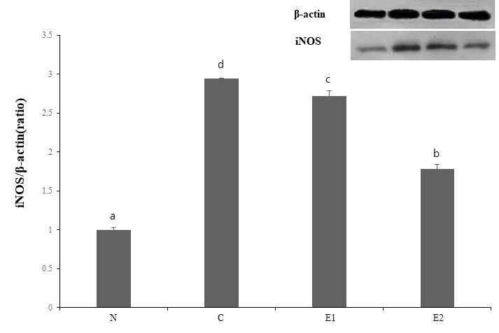
RAW 264.7 cell을 6-well plate에 1×10⁶ cells/mL로 분주 한 후 시험물질을 처치하여(100 및 200 µg/mL) 24시간 배양하였다. PBS로 2번 세척 후 RIPA buffer 200 μL/well 씩 처치하였으며 scraper를 이용하여 세포를 떨어뜨린 후 1.75 mL tube로 옮겨 10분간 원심분리 하여 단백질 농도를 정량하였다. 전기영동을 통해 iNOS를 정량하여 LPS 처치군과 시험물질 처치군간의 발현량을 비교 분석하였다. Band의 시각화는 ECL(Amersham)을 이용하였고 X-ray film에 가시화 하였다.
6. In vivo 소양감 억제 평가
생후 6주령의 수컷 ICR mouse를 ㈜대한바이오링크(충북 음성, 한국)에서 분양 받아 1주간 온도는 22±3℃, 습도는 50±5%, 명암주기 12시간으로 조절되는 표준사육 환경에서 적응한 뒤 실험에 사용하였다. 실험기간 동안 사료(Purina, 한국)와 물은 자유롭게 공급하였다. 소양감 유도 전 정확한 투여를 위해 전기제모기(electric clipper)를 이용하여 제모를 하였다.
모든 동물 실험 절차는 계명대학교 동물실험윤리위원회(IACUC)의 승인(승인번호. KM-2018-004)을 얻은 후 가이드라인에 의거하여 수행하였다. 소양감 억제 실험을 위해 각 군당 5마리씩 분류하였으며 정상군(N), Compound 48/80을 투여하여 소양감을 유도한 대조군(C), 소양감 유도후 Azelastine HCl을 처치한 양성 대조군(PC), 한약복합추출물(NI-01)을 처치한 실험군(T)으로 구성하였다(Table 2). 소양감 억제를 평가하기 위해 시험 물질은 각 용량당 10 mL/kg씩 2주간 동일한 시간에 경구 투여하였다. 마지막 경구투여 1시간 후 전기제모기(electric clipper)를 이용하여 마우스의 등 부위를 제모하고 compound 48/80을 saline에 1 mg/mL으로 조제하여 100 μL씩 제모된 등 부위에 피내 주사하여 소양감을 유도하였다. 정상군(N)은 compound 48/80의 용매물질(saline)을 피내 주사하였다.
소양감 유도에 따른 행동학적 특징을 관찰하기 위해 마우스가 뒷다리를 이용하여 목, 안면, 등 부위의 피부를 긁는 횟수를 기준으로 하였다. 또한, 마우스는 1초 동안 여러 차례 소파 행위를 하므로 한 번에 연속적으로 긁는 행위는 한번으로 간주하여 측정하였다(Kim and Kim 2007). Compound 48/80을 피내 주사한 후 30분간 촬영 및 측정하여 군간 비교하였다.
7. BCOP를 이용한 자극성 평가
자극 평가는 OECD guideline 43725)에 의거한 안점막자극 대체시험법(Bovine cornea opacity and permeability, BCOP)을 이용하여 측정하였다. 소 안구를 도축장에서 받아 각막을 분리하고 홀더에 장착 후 MEM(minimum essential medium, with out phenol red)을 양쪽 chamber에 채워 32℃에서 1시간 전배양 하였다. 전배양 후 opacitometer로 초기 혼탁도를 측정한 후 홀더의 상단 챔버의 MEM을 제거하고, 시료 750 µL를 각막 상피에 처치한 후 10분간 배양하였다. 시료 처치 후, MEM(with phenol red)으로 3회 세척하고 최종적으로는 MEM(with out phenol red)으로 세척하였다. 각막을 세척 후, MEM(with out phenol red)을 채워 2시간간 후배양 하였으며, 후배양 후 opacitometer를 이용하여 최종 혼탁도를 측정하였다. 후기 혼탁도 측정 완료 후 32℃로 예열하여 준비한 fluorescein sodium을 각막 상피에 1 mL씩 도포하고 32℃에서 90분간 incubation하였다. 배양 종료 후 하단 챔버 내 MEM은 균일하게 혼합하여 96-well plate에 분주 후 spectrophotometer를 사용하여 490 nm에서 흡광도를 측정하였다.
7. 통계처리
실험 결과는 평균값과 표준편차로 나타내었으며, 통계 분석은 SPSS 21.0 for windows program(SPSS Inc, USA)을 이용하여 일원배치분산분석(one-way ANOVA)을 실시하였다. 각 그룹 간의 차이를 검정하기 위한 사후분석은 Duncan's multiple range test를 실시하였으며 p<0.05, p<0.01, p<0.001에서 유의성을 검증하였다.
결 과
1. Antioxidant activity of NI-01
항산화 활성 평가를 위해 DPPH 전자공여능을 측정한 결과 한약복합추출물(NI-01)은 농도 의존적으로 활성이 증가되는 양상을 나타냈고, 250 μg/mL에서 70%이상의 활성을 보였으며 500 μg/mL와 1000 μg/mL에서는 양성대조군인 ascorbic acid와 동일 농도에서 유사한 높은 전자공여능을 나타냈다. 전자공여능에 대한 NI-01의 SC50은 127.3 μg/mL으로 산출되었다. ABTS+ radical 소거능의 경우에도 시험물질은 농도 의존적으로 증가하는 양상을 나타냈으며 250 μg/mL에서 90% 이상의 활성을 보였다. 또한, 500과 1000 μg/mL 농도에서 NI-01은 ascobic acid와 유사하게 높은 활성을 나타냈다. ABTS+ radical 소거능에 대한 NI-01의 SC50은 114.4 μg/mL으로 산출되었다(Fig. 1).
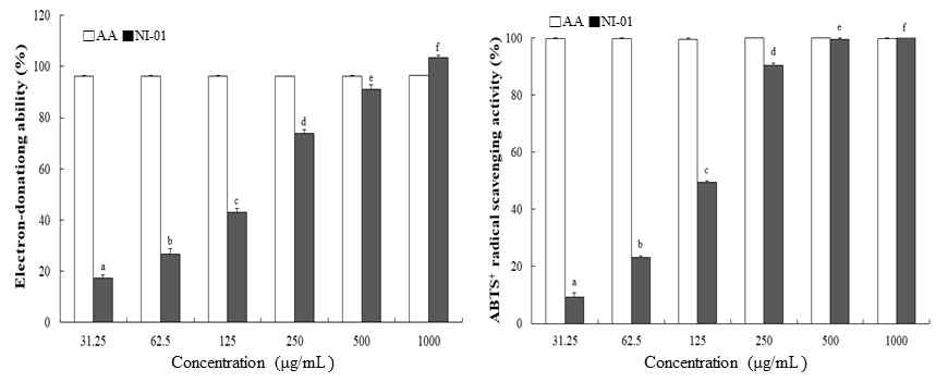
Electron-donating ability and ABTS+ radical scavenging activity of NI-01. Values represent mean ± SD of three independent experiments. Values with different letters are significantly different(p<0.05) by ANOVA and Duncan’s multiple range test. AA : Ascorbic acid. NI-01: Water extract of six herbal complex.
2. RAW 264.7 cell에서의 Nitric Oxide inhibitory rate
LPS만 처치한 후 세포 생존율은 63.8%로 나타났으며 NI-01의 전 농도에서 70% 이상의 세포생존율을 확인하였다. LPS와 NI-01을 같이 처치하였을 때 세포생존율은 70% 이상으로 개선되었고, 이는 세포독성에 영향이 적은 농도로 판단되어, 100 µg/mL 이하 농도에서 NO생성 저해효과 실험을 진행하였다.
NO생성 저해에 대한 한약복합추출물(NI-01)의 IC50은 20.54 μg/mL으로 산출되었으며 농도 의존적인 양상으로 NO 생성을 저해하였다. 또한, 50과 100 µg/mL 농도에서 각각 14.9%, 4.5%의 낮은 NO 생성을 나타냈다(Fig. 2).
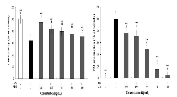
Effects of NI-01 on cell viability and NO production in LPS-stimulated RAW 264.7 cells. Control: LPS-treatment control. The values are mean±SD of 3 independent measurements. Values with different letters are significantly different (p<0.05) by ANOVA and Duncan’s multiple range test. ***p<0.001 as compared to the LPS only control group by ANOVA and Duncan’s test. NI-01: Water extract of six herbal complex.
3. RAW 264.7 cell에서의 iNOS 단백질 정량
한약복합추출물(NI-01) 100 및 200 µg/mL을 처치한 RAW 264.7 cell에서 LPS만 처치한 군에 비해 iNOS의 발현이 감소하였으며 특히 200 μg/mL 농도에서 현저하게 감소하였음을 확인하였다(Fig. 3).
4. In vivo 소양감 억제 평가 결과
시료 처치 및 소양감을 유도한 후 30분간 촬영하여 마우스가 뒷다리를 이용하여 목, 안면, 등 부위의 피부를 긁는 횟수를 측정하였을 때, C군과 PC군은 각각 159.0±35.9회, 39.9±27.1회의 긁는 횟수를 나타냈다. 한약복합추출물(NI-01)을 처치한 실험군의 긁는 횟수는 농도 의존적으로 감소하는 양상으로 나타났다. 특히 T3군은 PC군과 유사한 높은 소양감 억제를 나타냈으며, 소양감 억제에 대한 한약복합추출물(NI-01)의 IC50은 135.3 mg/kg으로 산출되었다(Fig. 4).
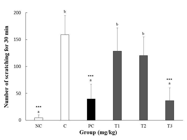
Inhibitory effect of orally administered NI-01 and Azelastine HCl on Compound 48/80 induced scratching behavior. Values are mean±SD of 5 mice. Test compounds were orally administered to ICR mice for 2 weeks. NC: DW+saline, C: DW+C48/80, PC: Azelastine HCl(20 mg/mL)+C48/80, T1: NI-01 40 mg/mL+C48/80, T2: NI-01 80 mg/mL+C48/80, T3: NI-01 160 mg/mL+C48/80. Values with different letters are significantly different (p<0.05) by ANOVA and Duncan’s multiple range test. ***p<0.001 as compared to the C48/80 only control group by ANOVA and Duncan’s test. NI-01: Water extract of six herbal complex.
5. Evaluation on eye irritation by BCOP assay
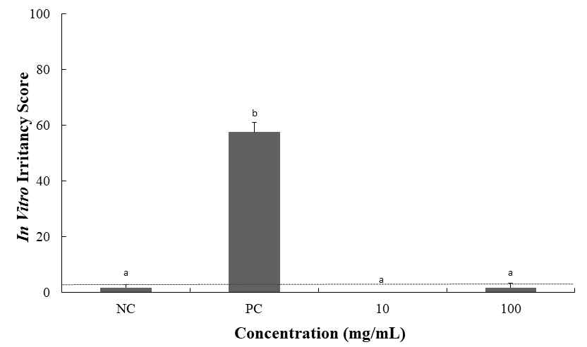
안자극지수(IVIS)는 혼탁도와 투과도를 이용하여 산출하였으며, 시험 결과 음성대조군의 IVIS 값은 1.6으로 비 자극, 양성대조군은 57.6으로 자극물질로 판정하였다. 한약복합추출물(NI-01)도 모든 농도에서 3 이하의 값을 보여 비 자극물질로 분류되었다(Fig. 5).
고 찰
아토피 피부염은 난치성 피부질환으로 유병율이 지난 30년간 급증하고 있는데, 이는 대기오염의 증가와 함께 주거환경의 변화로 인한 항원의 노출 증가, 모유수유 감소 등의 영향 때문일 것으로 추측되고 있다. 아토피 피부염 환자들은 심한 소양감과 염증반응으로 인해 만성 피로를 겪으며 학업뿐만 아니라 신체적, 사회적 및 심리적 문제에 부딪히게 되며 이에 따른 경제적 손실이 증가함에 따라 아토피 피부염에 대한 치료에 관심이 증가하고 있는 추세이다26). 현재 한의학에서 한약재를 단독으로 사용하는 경우가 드물며 다양한 한약재를 혼합하여 사용하고 있는 추세이므로 본 연구에서도 한약복합추출물(NI-01)을 이용한 In vivo 및 In vitro 연구를 통해 항산화, 항염증, 소양감 억제 효과를 확인하여 아토피 개선 기능성 소재로서의 가능성을 알아보고자 하였다.
일반적으로 한약추출물의 경우, 항산화능이 우수한 물질을 선별하여 다양한 효능평가에 활용하고 있는데, 목단피 추출물27)이나 오미자 추출물19)의 경우 높은 항산화능 물질로 보고되고 있다. 본 시험에 사용된 한약복합추출물(NI-01)도 DPPH assay와 ABTS+ assay에서 우수한 항산화능을 확인할 수 있었다.
항염증 평가를 위해 RAW 264.7 cells에 LPS를 처치하여 NO 생성을 유도한 후 한약복합추출물(NI-01) 처치에 따른 저해효과를 평가하였다. RAW 264.7 cells는 항염증 약물의 효능평가에 많이 사용되며, LPS는 세포를 자극하여 염증 반응을 유도하여 NO, PGE2와 같은 염증 매개물질 생성을 촉진하는 물질로 사용된다28,29). 한약복합추출물(NI-01) 100 µg/mL의 세포생존율은 LPS 처치하였을 때 보다 높게 나타나 LPS에 의한 세포독성을 완화시켜주는 역할을 한 것으로 생각되며, 세포에 미치는 NI-01 독성에 대한 IC50은 1136 μg/mL으로 산출되었다. 한약복합추출물(NI-01)의 NO생성 저해에 대한 IC50은 20.54 μg/mL으로 산출되었으며 농도 의존적으로 높은 항염증 효과를 확인할 수 있었다.
계지 추출물은 LPS 처리로 인한 NO와 PGE2 생성을 억제할 뿐만 아니라 iNOS 및 COX-2의 발현을 저해시켜 항염 효과가 있는 것으로 알려져 있으며14), 금은화는 NFκB가 핵 안으로 전위(translocation)됨을 억제하여 NO, iNOS, TNF-α, COX-2, IL-6, IL-1β의 감소시킴으로 항염증 효과를 발생시키는 것으로 보고되고 있다30,31). 이외에도 향유 추출물은 IL-6, IL-1α, COX-2, NO 생성 억제에 효과적이며32), 목단피와 도인 혼합추출물의 경우 목단피 단독 추출물에 비해 효과적으로 NO 생성을 저해하는 것으로 보고되어 있다33). 이는 목단피가 다른 약재와 혼합할 경우 상승 작용에 기인하는 것을 의미한다. 본 시험에서도 NI-01을 구성하는 시료 중에 계지, 금은화, 향유, 목단피가 항염증 효과에 크게 작용하여 나타났다. 특히, 항산화에 효과적인 목단피가 다른 시료와의 상승 작용으로 인해 높은 항염증 활성을 나타낸 것으로 사료된다. 항염 효능평가에 있어서도, NO 생성과 iNOS 단백질 발현이 시험물질에 의해 모두 감소하는 결과를 보였다. iNOS(nitric oxide synthases, inducible isoform)는 염증반응이 일어났을 때, 염증을 제거하기 위해 대식세포에 의해 발현되며 이때 iNOS에 의해 NO의 생성도 증가되게 되는데, 이들의 감소는 좋은 항염증 효과를 보일 수 있음을 암시한다.34)
제 1형 알러지성 염증 반응에 해당되는 아토피 피부염의 주 증상은 가려움증으로 피부를 긁는 행위로 인해 2차 피부질환을 유발하게 되며 본 연구에서 한약복합추출물(NI-01)의 소양감 억제 효능을 확인하였다. 소양증 연구에 대한 마우스 모델에서 사용하는 가려움증제는 substancs P, histamine, compound 48/80 등이 있으며35), 그 중 compound 48/80은 비만세포의 세포막에 작용하여 세포 외에 존재하는 칼슘을 세포내로 유입시키고 cyclic adenosine monophosphate(cAMP)와 cyclic guanosine-3’,5’ monophosphate(cGMP)양을 변화시켜 비만세포의 탈 과립으로 히스타민이 유리되어 소양감을 유도시키는 역할을 한다36,37). 정상군과 compound 48/80을 유도한 군의 긁는 횟수는 현저하게 높은 차이를 나타냈으며 한약복합추출물(NI-01)은 농도 의존적인 양상을 보였다. 한약복합추출물(NI-01) 160 mg/kg 처치 시 Azelastine HCl를 투여한 양성대조군과 유사한 수치로 높은 소양감 억제를 나타냈다.
우방자는 세포내 cAMP양을 증가시키는 비만세포 안정화를 통해 비만세포의 탈과립 및 혈장 histamine 유리를 감소시켜 compound 48/80에 의한 가려움증을 억제하며1), 목단피 추출물의 대표 성분 중 paeonol은 비만 세포와 RBL-2H3 세포에서 각각 히스타민과 TNF-α 방출을 억제하여 항 알레르기 활동에 기여한다고 알려져 있는데38), 본 시험에서 한약복합추출물(N1-01)은 목단피와 다른 시료 간의 상가 작용으로 더 좋은 소양감 억제 효과를 보여주고 있다. 또한 한약복합추출물(N1-01)은 안점막 자극 대체시험법으로 널리 알려져 있는 BCOP 시험법에서 비자극 물질로 판정되어 국소적인 자극에 있어서 안전성을 확인하였다.
본 연구의 결과를 종합해 볼때, 한약복합추출물(NI-01)은 높은 항산화 및 항염증 효과를 보였고 우수한 소양감 억제 효과를 나타냈으며 안점막 자극 시험에서 비 자극으로 판정되어 건강기능식품, 화장품 분야에서의 활용 가능성을 확인할 수 있었다. 최근 천연 소재를 이용한 기능성 소재 탐색 연구가 활발하게 이루어지고 있기 때문에, 본 시험에 사용된 다양한 복합 성분들에 대한 효능의 기전적 규명 및 허가 등을 위한 추가적 시험이 요구되어지지만 한약재 혼합사용을 통해 항염증 및 소양감 억제 등의 효능이 증가된 새로운 아토피 소재로서의 활용을 위한 기초자료를 제시할 수 있을 것이라고 판단된다.
References
- Kim SH, Lee JY, Kim DG. Effects of mori folium, arctii fructus, Lithospermum Erythrorhizon on the antiallergic response. J. Kyung Hee Univ. Med. Cent. 2005;21:71-9.
-
Kim GW, Bak JW, Sim BY, Kim DH. An experimental study for the development of prescription on atopic dermatitis. Kor. J. Herbology 2014;29:13-20.
[https://doi.org/10.6116/kjh.2014.29.4.13]

- Kim SA, Kang YH. Changes in ceramide in stratum corneum and anti-inflammatory effects of Sopungdojeok-tang on atopic dermatitis. Kor. J. Intern. Med. 2006;27:72-83.
-
Jeong YS, Jung HK, Youn KS, Kim MO, Hong JH. Physiological activities of the hot water extract from Eriobotrya japonica Lindl. J. Kor. Soc. Food Sci. Nutr. 2009;38:977-82.
[https://doi.org/10.3746/jkfn.2009.38.8.977]

- Yamashita H, Makino T, Inagaki N, Nose M, Mizukami H. Assessment of relief from pruritus due to Kampo medicines by using murine models of atopic dermatitis. J. Trad. Med. 2013;30:114-23.
-
Grewe M, Bruijnzeel Koomen CA, Schöpf E, Thepen T, Langeveld Wildschut AG, Ruzicka T. Krutmann J. A role for Th1 and Th2 cells in the immunopathogenesis of atopic dermatitis. Immunol. today 1998;19: 359-61.
[https://doi.org/10.1016/S0167-5699(98)01285-7]

-
Lee MH, Han MH, Yoon JJ, Song MK, Kim MJ, Hong SH, Choi YH. Medicinal Herb Extracts Attenuate 1-Chloro-2,4dinitrobenzene -induced Development of Atopic Dermatitis-like Skin Lesions. J. Life Sci. 2014;24:851-9.
[https://doi.org/10.5352/JLS.2014.24.8.851]

- Baek S, Choi JH, Ko SH, Lee YJ, Cha DS, Park EY, Jeon H. Antioxidant and Anti-inflammatory Effect of Nardostachys Chinensis in IFN-γ/LPS-stimulated Peritoneal Macrophage. J. Physiol. 2009;23:853-9.
- Kim HH, Park GH, Park KS, Lee JY, An BJ. Anti-oxidant and Anti-inflammation Activity of Fractions from Aster glehni Fr. Schm. Microbiol. Biotechnol. Lett. 2010;38:434-41.
-
Kim CH, Lee MA, Kim TW, Jang JY, Kim HJ. Anti-inflammatory effect of Allium hookeri root methanol extract in LPS-induced RAW264. 7 cells. J. Kor. Soc. Food Sci. Nutr. 2012;41:1645-8.
[https://doi.org/10.3746/jkfn.2012.41.11.1645]

-
Shen CZ, Park JK, Kim CG, Chun HJ, Kim IK. The anti-histamine effect of water soluble alkaloids extracted from Solanum nigrum L. Anal. Sci. Technol. 2016;29:186-93.
[https://doi.org/10.5806/AST.2016.29.4.186]

- Kim KS, Kim YB. The latest study tendency in mouse model of skin pruritus-Mainly on scratching behavior model. J. Kor. Med. Ophthalmol. Otolaryngol. Dermatol. 2007;20:142-50.
- Kweon MH, An HJ, Shin KS, Sung HC, Yang KS. Purification of complement system-activating polysaccharide from hot water extract of young stems of Cinnamomum cassia Blume. J. Food Sci. Technol. 1997;29:1-8.
- Park HJ, Lee JS, Lee JD, Kim NJ, Pyo JH, Kang JM, Lim S. The anti-inflammatory effect of Cinnamomi Ramulus. J. Kor. Oriental. Med. 2005;26:140-51.
- Bae JH, Kim MS, Kang EH. Antimicrobial effect of Lonicerae Flos extracts on food-borne pathogens. J. Food Sci. Technol. 2005;37:642-7.
-
Lee SJ, Lee MJ, Jung JE, Kim HJ, Bose S. In vitro Profiling of Bacterial Influence and Herbal Applications of Lonicerae Flos on the Permeability of Intestinal Epithelial Cells. Kor. Soc. Food Sci. Nutr. 2012;41:881-7.
[https://doi.org/10.3746/jkfn.2012.41.7.881]

-
Hwang JS, Han YS. Isolation and identification of antimicrobial compound from Mokdan bark (Paeonia suffruticosa ANDR). J. Kor. Soc. Food Sci. Nutr. 2003;32:1059-65.
[https://doi.org/10.3746/jkfn.2003.32.7.1059]

- Park SM, Jun DW, Park CH, Jang JS, Park SK, Ko BS, Choi SB. Hypoglycemic effects of crude extracts of Moutan Radicis Cortex. J. Food Sci. Technol. 2004;36:472-7.
- Kim JS, Choi SY. Physicochemical properties and antioxidative activities of Omija (Schzandra chinesis Bailon). Kor. J. Food Nutr. 2008;21:35-42.
- Seong KH, Kim JY, Lee JH, Park SK, Cho YH. Arctii Fructus is a Prominent Dietary Source of Linoleic Acid for Reversing Epidermal Hyperproliferation of Guinea Pigs. J. Nutr. 2003;36:819-27.
- Jeong JH, Lim HB. Chemical Composition and Biological Activities of Elsholtzia ciliata (Thunb.) Hylander. Kor. J. Medicinal Crop Sci. 2004;12:463-72.
- Youn KS, Hong JH, Kwon JH, Choi YH. Optimization for Elsholtzia ciliata Hylander extraction using supercritical carbon dioxide. Kor. J. Food Preserv. 2006;13:363-8.
-
Blois MS. Antioxidant determinations by the use of a stable free radical. Nature 1958;181:1199-200.
[https://doi.org/10.1038/1811199a0]

-
Re R, Pellegrini N, Proteggente A, Pannala A, Yang M, Rice-Evans C. Antioxidant activity applying an improved ABTS radical cation decolorization assay. Free Radic. Biol. Med. 1999;26:1231-7.
[https://doi.org/10.1016/S0891-5849(98)00315-3]

- OECD. OECD guideline for the testing of chemicals, NO. 437: Bovine Corneal Opacity and Permeability Test Method for Identifying i) Chemicals Inducing Serious Eye Damage and ii) Chemicals Not Requiring Classification for Eye Irritation or Serious Eye Damage. 2017.
-
Leung DY, Boguniewicz M, Howell MD, Nomura I, Hamid QA. New insights into atopic dermatitis. J. Clin. Invest. 2004;113:651-7.
[https://doi.org/10.1172/JCI21060]

-
You JK, Chung MJ, Kim DJ, Seo DJ, Park JH, Kim TW, Choe M. Antioxidant and tyrosinase inhibitory effects of Paeonia suffruticosa water extract. J. Kor. Soc. Food Sci. Nutr. 2009;38:292-6.
[https://doi.org/10.3746/jkfn.2009.38.3.292]

- Kim TY. Effect of Gagam-Danguieumja through regulation of MAPK on LPS-induced inflammation in RAW 264.7 cells. Kor. J. Intern. Med. 2013;34:339-48.
-
Han MH, Lee MH, Hong SH, Choi YH, Moon JS, Song MK, Hwang HJ. Comparison of anti-inflammatory activities among ethanol extracts of Sophora flavescens, Glycyrrhiza uralensis and Dictamnus dasycarpus, and their mixtures in RAW 246.7 murine macrophages. J. Life Sci. 2014;24:329-35.
[https://doi.org/10.5352/JLS.2014.24.3.329]

- Lee DE, Lee JR, Kim YW, Kwon YK, Byun SH, Shin SW, Suh SI, Kwon TK, Byun JS, Kim SC. Inhibition of Lipopolysaccharide-Inducible Nitric Oxide Synthase, TNF-α, IL-1β and COX-2 Expression by Flower and Whole Plant of Lonicera japonica. Korean J. Oriental Physiol. Pathol. 2005;19:481-9.
-
Cha HY, Jeong AR, Cheon JH, Ahn SH, Park SY, Kim KB. The Anti-oxidative and Anti-inflammatory Effect of Lonicera Japonica on Ulcerative Colitis Induced by Dextran Sulfate Sodium in Mice. J. Pediatr. 2015;29:54-64.
[https://doi.org/10.7778/jpkm.2015.29.3.054]

- Lee DW, Kim YJ, Kim YS, Eom SY, Kim JH. Inhibitory Effect of Melanogenesis and Anti-inflammatory Effect of Elsholtzia ciliata Extract and Its Application as a Cosmeceutical Ingredient. J. Soc. Cosmet. Scientists Korea 2006;32:219-25.
- Kim YI, Lee SJ, Huh J, Lee TH, Shin DG, Lee JC, Yun YG. Effect of Water Extract of Peonia Suffruticosa and Prnus Percica on Anti-inflammation. Herbal Formula Science 2010;18:105-20.
-
Fugen A. iNOS-mediated nitric oxide production and its regulation. Life Sci. 2004;75:639-53.
[https://doi.org/10.1016/j.lfs.2003.10.042]

-
Jang SE, Ryu KR, Park SH, Chung S, Teruya Y, Han MJ, Kim DH. Nobiletin and tangeretin ameliorate scratching behavior in mice by inhibiting the action of histamine and the activation of NF-κB, AP-1 and p38. Int. Immunopharmacol. 2013;17:502-7.
[https://doi.org/10.1016/j.intimp.2013.07.012]

-
Rothschild AM. Mechanisms of histamine release by compound 48/80. Br. J. Pharmacol. 1970;38:253-62.
[https://doi.org/10.1111/j.1476-5381.1970.tb10354.x]

-
Li GZ, Chai OH, Song CH. Inhibitory Effect of Rubus Coreanus on Compound 48/80 or Anti-DNP IgE-Induced Mast Cell Activation. Nat. Immunol. 2004;4:100-7.
[https://doi.org/10.4110/in.2004.4.2.100]

-
Jiang S, Nakano Y, Yatsuzuka R, Ono R, Kamei C. Inhibitory effects of Moutan cortex on immediate allergic reactions. Biol. Pharm. Bull. 2007;30:1707-10.
[https://doi.org/10.1248/bpb.30.1707]


