Balb/c 생쥐에 대한 어싱 매트리스에 의한 항염 효과
Ⓒ The Society of Pathology in Korean Medicine, The Physiological Society of Korean Medicine
Abstract
Earthing, caused by direct skin contact with the Earth's surface, is used to reduce the symptoms of inflammation (fever, fever, swelling and pain). However, there is little evidence to support the anti-inflammatory effects of earthing mattresses. Therefore, this study was conducted to investigate whether anti-inflammatory effect of earthing mattress using an in vivo animal model. The anti - inflammatory effect was evaluated by measuring ear thickness and foot volume in 12-O-tetradecanoylphorbol-13 acetate (TPA) - induced ear edema and carrageenan - induced paw edema model, respectively. Balb/c mouse in carrageenan paw edema model showed significant anti - inflammatory effect in the group treated with earthing mattress for 4 hours or 24 hours for 3 days. For females, the anti-inflammatory effect was greater when the earthing mattress was added to the mattress than the mattress alone treatment. From the above results, it was found that the female responds more to the effect of the earthing as well as the mattress effect. In addition, when the male and female Balb/c mice were exposed to mattresses and earthing mattresses for 24 h for 3 days, respectively, the mattress and earthing mattresses showed significant inhibition of IL (Interleukin)-1β levels compared to the control. In the TPA ear edema model, Balb/c mouse showed significant anti - inflammatory effect in the group treated with the earthing mattress for 4 hours or 24 hours for 3 days. Both males and females showed more anti-inflammatory effects when they were exposed to earthing mattresses with mattresses added to the mattresses. From the above results, it was found that both male and female respond to the effect of earthing as well as the mattress effect in the TPA ear edema model. In conclusion, in this study, we have verified that earthing mattress shows inhibitory effects on TPA and carrageenan-induced inflammation. From these results, it is suggested that the anti-inflammatory effect can be expected by applying the earthing mattress to patients suffering from inflammatory diseases. However, there is a need to pinpoint exactly how the earthing mattress relieves inflammation, and further research is needed to investigate the mechanism.
Keywords:
Earthing, Anti-inflammation, Carrageenan, 12-O-tetradecanoylphorbol-13 acetate (TPA), Cytokine서 론
염증은 유해 물질에 대한 신체의 자기 보호의 반응이며 유해한 자극을 제거하고 치유과정을 시작하기 위한 유기체의 보호조치 이다1). 이러한 염증은 반응의 기간에 따라 급성 또는 만성으로 분류되며 급성 염증 반응은 혈장 및 백혈구의 손상 또는 감염 부에 침투하여 시작되며2) 톨 유사 수용체 (Toll-like receptor) 등 면역 계통의 수용체에 의해서 유발될 수 있다3). 반면 만성 염증은 지속적인 자극에 대한 조절 불량 반응이며 조직의 오작동과 관련이 있다. 여기에는 암, 알레르기, 관절염, 즉 상 동맥경화증, 자가 면역 질환을 비롯한 만성 질환과 관련되어 있다2).
어싱 (Earthing)은 맨발이나 손과 같이 지구 표면과의 직접적인 피부 접촉 또는 다양한 접지 시스템을 의미하며 이러한 어싱은 수면 중 야간에 몸을 접지하면 아침 피로감, 주간 에너지 및 야간 통증 수준에서 긍정적인 영향을 미치는 것으로 나타났으며4) 어싱은 부상 후 홍역, 염증, 열, 부기, 통증 및 기능 상실의 염증 징후를 감소시키거나 예방한다고 알려져 있다5).
카라기난 (Carrageenan, Carr)은 해조류에서 추출한 고 분자량의 황산화 다당류를 지칭하며 유아용 조제유, 유제품, 아몬드 우유, 가공육 및 콩기름 제품과 같은 우유 대체 식품을 포함한 상업용 식품의 질감을 개선하기 위한 농축 및 유화제 식품 첨가물로 일반적으로 사용되는 이름이다6). 가장 일반적인 형태로 람다 (λ), 카파 (κ) 및 이오타 (ι)가 있다7). 카라기난은 황산화 이당류의 조성을 가지고 있어 자연적으로 발생하는 글리코사미글리칸 (Glycosaminoglycans)을 닮았다7).
카라기난은 재현성 염증 반응을 일으키며 항염증제의 급성 염증 및 효과를 검사하기 위한 표준 화학 물질로 남아 있다8). 카라기난은 발의 염증을 유도하여 동물의 항염증 반응을 선별하는 데 흔히 사용되는 기법이다9). 랫트 (Rat) 뒷다리의 카라기난 유발 부종은 광범위하게 사용된 염증 모델이다10). 이 염증 작용제는 히스타민 (Histamine), 세로토닌 (5-Hydroxytryptamine, 5-HT), 프로스타글란딘 (Prostaglandin, PG)을 유도하며 염증을 일으켜서 실험 약학에서 흔히 사용되며 동물에서 염증의 장 모델로 사용되고 흉막 공간 및 복막을 포함한 Extra-colonic sites에 염증을 유발한다11-14).
12-O-tetradecanoylphorbol-13 acetate (TPA)는 Keratinocytes에서 Proinflammatory cytokines의 발현을 유도하는 것으로 밝혀졌으며 피부 염증에 관여하는 것으로 밝혀져 있다15). 귀 부종은 치료제의 항염증 활성을 평가하기 위한 유용한 약리학적 모델로 널리 사용됐으며16,17), Phorbol ester 중 하나인 TPA는 부종, 표피 과형성 및 염증 매개체의 과생산을 나타내며 피부 염증을 유발할 수 있다16,18). TPA에 의해 유발된 마우스 귀 부종은 항염증제 활성을 테스트하기 위한 동물 모델로 사용되었습니다. 이 모델에서 피부 부종은 프로스타글란딘 E2 (Prostaglandin E2, PGE2)와 류코트리엔 B4 (Leukotriene B4, LTB4)와 같은 에이코사노이드 (Eicosanoid)의 함량 증가와 관련이 있다고 알려져 있다19).
따라서 본 연구진들은 어싱 매트리스의 항염증 유효성을 평가하기 위하여 재현성 염증 반응을 일으키며 항염증제의 급성 염증 및 효과를 검사하기 위한 표준 화학 물질인 카라기난과 Keratinocytes에서 Pro-inflammatory cytokines의 발현을 유도하는 TPA를 사용하여 in vivo 동물 모델에 염증을 유발 후 어싱 매트리스를 노출해 항염증 유효성을 평가하였다.
재료 및 방법
1. 재료
Walking Test Set EFM 023 (BGT UAC-110)은 Kleinwachter GmbH사에서 구매하였다. Mouse IL-1β/IL-1F2 kit는 R&D Systems (Abingdon, UK) 사에서 구매하였고, TPA (P1585), 카라기난 (22049) 등은 시그마 (St. Louis, MO, U.S.A.) 사에서 구매하여 사용하였다. 어싱 매트리스는 닥터프렌드(Korea)의 어싱 매트리스를 사용했으며 수면 매트리스 사이에 신체 내의 전위차를 제거하는 판을 넣어 매트리스 형식으로 제작하여 만든 소재를 이용하였다.
2. 실험 동물
Balb/c mouse (생후 5주령)을 오리엔트 바이오로부터 구매하고, 온도 20±2도, 습도 50±10%, 12시간 명암주기가 유지되는 동물실에서 일주일 동안 예비 사육하였다. 어싱 매트리스에 4h 3일, 5일, 24h 3일, 5일 노출한 후에 다음의 염증 실험을 실시하였다. 대조군은 일반 스펀지로 진행하고, 처리군은 어싱 소재의 매트리스로 진행하였다.
3. 카라기난 염증 모델에서 항염 효능 평가
발바닥 부종 염증을 유도하기 위해 1% 람다 카라기난을 50ul씩 오른쪽 발바닥에 도포하고 카라기난 처리 4시간 후 Ketamine (100mg/kg), Xylazine (10mg/kg)로 마취하였다. Vernier calipers로 발의 두께와 발바닥 부종의 두께 변화를 좌우 앞발 사이에서 측정하였다.
4. 뒤 염증 모델에서 항염 효능 평가
귀 부종 염증을 유도하기 위해 오른쪽 귀에 아세톤으로 녹인 TPA 2ug/20ul/ear씩 도포하였다. TPA 처리 4시간 후 Ketamine (100mg/kg), Xylazine (10mg/kg)로 마취 후 귀를 잘라내고 잘라낸 귀를 지름 6.5mm 펀치로 같은 크기의 조직 확보 후 Vernier calipers로 귀의 두께를 왼쪽 귀와 오른쪽 귀 사이의 밀리그램 차이로 측정하였다.
5. 어싱 매트리스 노출
Walking Test Set EFM 023 (BGT UAC-110) 인체 전위측정기를 통해 측정 전 229V가 어싱 매트리스에 발을 올린 후 0V로 내려간 것을 보고 어싱이 되었음을 확인 후 쥐를 어싱 매트리스에 시간별로 노출해 실험을 진행했다.
6. 사이토 카인 발현 측정
카라기난 주입 후 4시간 후에 피부 샘플을 채취했다. 시료는 10,000g x 15분 x 4℃으로 원심분리 후 RIPA buffer를 사용하여 조직 파열 장치로 균질화 시켰다. IL-1β 측정 키트 (R&D Systems, Abingdon, UK)를 사용하여 ELISA를 수행하였다. 흡광도는 450nm, 540nm에서 측정하였다. 결과는 조직의 g당 사이토 카인의 피코그램 (Pico gram, pg)으로 표현하였다.
7. 통계
개별 군의 평균을 % 대조군으로 전환하고 평균 ± S.E.M으로 나타냈다. 그룹 간 차이는 Post-hoc test로서 One-way ANOVA와 HolmeSidak test를 사용하여 평가하였고, p<0.05는 유의한 차이가 있는 것으로 간주하였다.
결 과
1. 어싱 매트리스의 3일간 시간별 노출에 따른 항염 효능
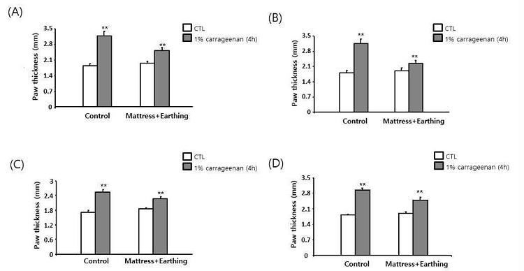
Balb/C 수컷 5마리씩 어싱 매트리스에 1h, 2h, 4h, 6h씩 3일간 노출시킨 후 카라기난 실험을 시행하여 대조군과 처리군의 염증 발현을 측정함으로써 어싱의 항염증 효과를 알아보았다.
Balb/C 수컷을 4h 3일 노출한 처리군이 1h, 2h, 6h씩 3일간 노출한 처리군보다 카라기난에 의해 유발된 염증을 유의하게 억제한다는 것을 관찰했다(Fig. 1). 이를 다시 확인하기 위하여 날짜별로 4h 3일, 5일로 날짜를 늘려 실험을 진행하였다.

Inflammation levels in the male mouse exposed to Earthing mattress for 1h/day, 2h/day, 4h/day, 6h/day for 3 days. Fig1A After exposed to Earthing mattress for 1h/day for 3 days. Fig 1B After exposed to Earthing mattress for 2h/day for 3 days. Fig 1C After exposed to Earthing mattress for 4h/day for 3 days. Fig 1D After exposed to Earthing mattress for 6h/day for 3 days. λ-Carrageenan at 1% was injected into the hindpaw, causing acute inflammation. Respectively, average of each group converted to % control and result represent the mean ± S.E.M. (n=5) ** p < 0.01 vs control.
2. 카라기난으로 유도된 염증 마우스 모델에서 어싱 매트리스 4시간 노출에 의한 항염 효능
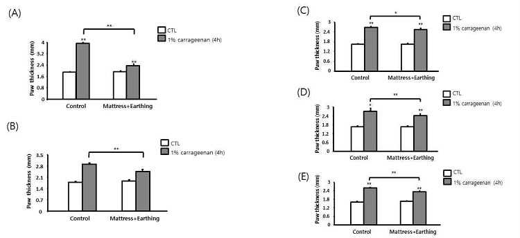
어싱의 항염증 효과를 알아보기 위해 4h 3일, 5일로 Balb/c 수컷 및 암컷 5마리씩 어싱 매트리스에 노출한 후 카라기난 실험을 수행하여 대조군과 처리군의 염증 발현을 측정하였다.
Balb/c mouse 수컷과 암컷 모두 4h 3일, 5일 처리한 어싱 매트리스 처리군이 대조군보다 카라기난에 의해 유발된 염증을 유의하게 억제한다는 것을 관찰했다. 그러나 암컷의 경우 억제 효과가 약하여 날짜를 7일로 늘려 실험을 진행하였다(Fig. 2). 이러한 결과는 어싱 매트리스가 카라기난에 의해 유도된 염증을 억제한다는 것을 보여준다. 이후 노출 시간을 24시간으로 늘려 추가실험을 진행하였다.

Inflammation levels in the Carrageenan injected mouse exposed to Earthing mattress for 4h/day. Anti-inflammation effect of Earthing mattress in the Carrageenan induced mouse inflammation. Fig 3A Male mouse was exposed to the Earthing mattress 4h/day for 3days. Fig 3B Male mouse was exposed to the Earthing mattress for 4h/day for 5 days. Fig 3C Female mouse was exposed to the Earthing mattress for 4h/day for 3days. Fig 4D Female mouse was exposed to the Earthing mattress for 4h/day for 5days. Fig 4E Female mouse was exposed to the Earthing mattress for 4h/day for 7days. After that, λ-Carrageenan at 1% was injected into the hindpaw, causing acute inflammation. Respectively, average of each group converted to % control and result represent the mean ± S.E.M (n=5) * p < 0.05 vs controls, ** p < 0.01 vs control.
3. 카라기난으로 유도된 염증 마우스 모델에서 어싱 매트리스 24시간 노출에 의한 항염 효능
어싱의 항염증 효과를 알아보기 위해 24시간 3일로 Balb/c 수컷 및 암컷 5마리씩 어싱 매트리스에 노출한 후 카라기난 실험을 수행하여 대조군과 처리군의 염증 발현을 측정하였다.
Balb/c mouse 수컷과 암컷을 24h 3일 처리한 어싱 매트리스 처리군이 대조군보다 카라기난에 의해 유발된 염증을 유의하게 억제한다는 것을 관찰했다(Fig. 3). 이러한 결과는 어싱 매트리스가 카라기난에 의해 유도된 염증을 억제한다는 것을 보여준다. 이런 결과가 어싱의 효능인지 아니면 매트리스의 효능인지를 확인하기 위하여, 그룹을 늘려 실험을 진행하였다.

Inflammation levels in Carrageenan injected mouse exposed to Earthing mattress for 24h/day. Fig 4A Male mouse was exposed to the Earthing mattress for 24h/day for 3 days. Fig 4B Female mouse was exposed to the Earthing mattress for 4h/day for 3 days. After that λ-Carrageenan at 1% was injected into the hindpaw, causing acute inflammation. Respectively, average of each group converted to % control and result represent the mean ± S.E.M. (n=5) ** p < 0.01 vs control.
4. 카라기난으로 유도된 염증 마우스 모델에서 어싱 매트리스 및 매트리스 24시간 노출에 의한 항염 효능
어싱의 항염증 효과를 알아보기 위해 24시간 3일로 Balb/c 수컷 및 암컷 5마리씩 어싱 매트리스에 노출한 후 카라기난 실험을 수행하여 대조군과 처리군의 염증 발현을 측정하였다.
Balb/c mouse 수컷과 암컷을 24h 3일 매트리스와 어싱 매트리스에 노출했을 때, 어싱 매트리스 노출군이 대조군보다 염증이 유의하게 억제된 것을 확인할 수 있다(Fig. 4). 또한 암컷을 노출한 어싱 매트리스 노출군에서 매트리스 노출군보다 확연하게 억제된 것을 관찰할 수 있다(Fig. 4). 또한 어싱 매트리스 24시간 노출에 의한 사이토 카인 변화를 확인하였다.

Inflammation levels in Carrageenan injected male mouse and female mouse exposed to mattress or Earthing mattress for 24h/day. Fig 5A Male mouse was exposed to the mattress or Earthing mattress for 4h/day for 3 days. Fig 5B Female mouse was exposed to the mattress or Earthing mattress for 4h/day for 3 days. After that λ-Carrageenan at 1% was injected into the hindpaw, causing acute inflammation. Respectively, average of each group converted to % control and result represent the mean ± S.E.M. (n=5) * p < 0.05 vs controls, ** p < 0.01 vs control.
5. 카라기난으로 유도된 염증 마우스 모델에서 어싱 매트리스 24시간 노출에 의한 사이토 카인 발현
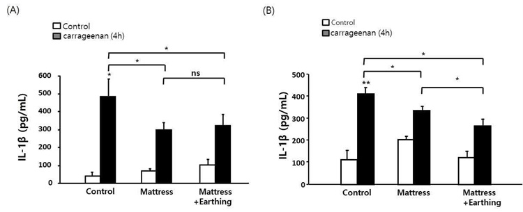
어싱의 항염증 효능을 확인하기 위해 24h 3일 Balb/c 수컷, 암컷 5마리씩 어싱 매트리스에 노출한 후 카라기난 실험을 수행하였다. 카라기난 주사 4시간 후 발 조직을 얻은 후 ELISA에 의해 IL-1β 수치를 측정하였다.
Balb/c mouse 수컷, 암컷을 24h 3일 매트리스와 어싱 매트리스에 각각 노출했을 때, 어싱 매트리스 노출군에서 대조군보다 유의하게 억제된 것을 확인할 수 있다(Fig. 5).

Changes of cytokine levels in the Carrageenan injected male mouse and female mouse exposed to Earthing mattress for 24h/day. for 3 days Fig 6A Inflammatory male mouse model induced to Carrageenan. Fig 6B Inflammatory female mouse model induced to Carrageenan. Respectively, after exposed to Earthing mattress for 24h/day for 3days, used to IL-1β kit. Average of each group converted to % control and result represent the mean ± S.E.M. (n=5) * p < 0.05 vs controls.
6. TAP로 유도된 염증 마우스 모델에서 어싱 매트리스 4시간 노출에 의한 항염 효능
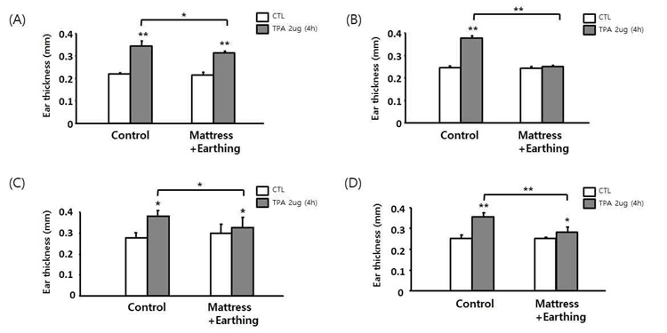
어싱의 항염증 효과를 알아보기 위해 4h 3일, 5일로 Balb/c 수컷 및 암컷 5마리씩 어싱 매트리스에 노출한 후 TPA 실험을 수행하여 대조군과 처리군의 염증 발현을 측정하였다.
Balb/c mouse 수컷과 암컷을 4h 3일, 5일 처리한 어싱 매트리스 처리군이 대조군보다 TPA에 의해 유발된 염증을 유의하게 억제한다는 것을 관찰했다(Fig. 6). 이러한 결과는 어싱 매트리스가 TPA에 의해 유도된 염증을 억제한다는 것을 보여준다. 또한 24h로 시간을 늘려 추가 실험을 진행하였다.

Inflammation levels in the TPA injected male mouse and female mouse exposed to Earthing mattress for 4h/day. Fig 8A Male mouse was exposed to the Earthing mattress for 4h/day for 3days. Fig 8B Male mouse was exposed to the Earthing mattress for 4h/day for 5days. Fig 8C Female mouse was exposed to the Earthing mattress for 4h/day for 3days. Fig 8D Female mouse was exposed to the Earthing mattress for 4h/day for 5days. Mouse was induced to ear edema on using TPA. Average of each group converted to % control and result represent the mean ± S.E.M. (n=5) * p < 0.05 vs controls, ** p < 0.01 vs control.
7. TPA로 유도된 염증 마우스 모델에서 어싱 매트리스 24시간 노출에 의한 항염 효능
어싱의 항염증 효과를 알아보기 위해 24시간 3일로 Balb/c 수컷 및 암컷 5마리씩 어싱 매트리스에 노출한 후 TPA 실험을 수행하여 대조군과 처리군의 염증 발현을 측정하였다.
Balb/c mouse 수컷과 암컷을 24h 3일 처리한 어싱 매트리스 처리군이 대조군보다 TPA에 의해 유발된 염증을 유의하게 억제한다는 것을 관찰했다(Fig. 7). 이러한 결과는 어싱 매트리스가 TPA에 의해 유도된 염증을 억제한다는 것을 보여준다. 이러한 결과가 어싱의 효능인지 아니면 매트리스의 효능인지 확인하기 위하여, 그룹을 늘려 실험을 진행하였다.

Inflammatory levels in the TPA injected male mouse and female mouse exposed to Earthing mattress for 24h/day. Fig 9A Male mouse was exposed to Earthing mattress for 24h/day for 3 days or 5 days. Fig 9B Female mouse was exposed to Earthing mattress for 24h/day for 3 days or 5 days. Mouse was induced to ear edema on using TPA. Average of each group converted to % control and result represent the mean ± S.E.M. (n=5) * p < 0.05 vs controls, ** p < 0.01 vs control.
8. TPA로 유도된 염증 마우스 모델에서 어싱 매트리스 24시간 노출에 의한 함염 효능
어싱의 항염증 효과를 알아보기 위해 24시간 3일로 Balb/c 수컷 및 암컷 4마리씩 어싱 매트리스에 노출한 후 TPA 실험을 수행하여 대조군과 처리군의 염증 발현을 측정하였다.
Balb/c mouse 수컷과 암컷을 24h 3일 매트리스와 어싱 매트리스에 노출했을 때, 어싱 매트리스 노출군이 대조군, 매트리스 노출군보다 염증이 유의하게 억제된 것을 확인할 수 있다(Fig. 8). 이를 통하여 어싱 매트리스가 염증 억제에 대해서 효과가 있음을 나타낸다.

Inflammatory levels in the TPA injected male mouse and female mouse exposed to mattress or Earthing mattress for 24h/day. Fig 10A Male mouse was exposed to the mattress or Earthing mattress for 24h/day for 3 days or 5 days. Fig 10B Female mouse was exposed to the mattress or Earthing mattress for 24h/day for 3 days or 5 days. Mouse was induced to ear edema on using TPA. Average of each group converted to % control and result represent the mean ± S.E.M. (n=5) * p < 0.05 vs controls, ** p < 0.01 vs control.
고 찰
한의학에서 자연계의 모든 사물은 음과 양의 두 가지 상반되는 개념을 형성하며 어떠한 사물도 내부에서는 음적인 면과 양적인 면의 사이에서 상호대립, 상호의존, 상호소장 및 상호전화를 형성하여 음과 양의 평형을 유지하며 음과 양의 조화가 깨지면 인체 내에서 질병으로 나타나게 된다.
어싱은 맨발이나 손과 같이 지구 표면과의 직접적인 피부 접촉 또는 다양한 접시 시스템을 의미하며 이러한 어싱에 의해 무수히 많은 질병이 치료된다고 보고되었는데, 통증, 염증, 순환장애, 피로 등에 개선 효능의 사례가 많이 보고되고 있다5). 또한 수면 중에 야간에 몸을 접지하면 아침 피로감, 주간 에너지 및 야간 통증 수준에 긍정적인 영향을 미치는 것으로 나타났다4). 이러한 지구와의 직접 접촉을 통하여 흡수된 자유 전자의 유입으로 활성산소 (Reactive oxygen species, ROS)를 중화하여 급성 및 만성 염증을 감소시키는 것으로 나타난다20). 이는 한의학의 음양 조화와 관련하여 과생성된 양전자를 자유 전자로 조화를 맞추어 신체의 균형을 맞추는 것으로 생각된다.
염증은 숙주의 방어에 중요한 역할을 하는 필수 생리적 과정이며21), 유해한 자극을 제거하고 더 많은 상처로부터 보호하며 치유 과정을 시작하고 손상된 조직을 정상적인 생리 기능으로 회복시키는 것이다. 이러한 염증은 반응의 기간에 따라 급성 또는 만성으로 분류된다. 급성 염증 반응은 혈장 및 백혈구의 손상 또는 감염 부위의 침투에 의해서 시작되며2), 만성 염증은 지속적인 유해 자극에 대한 조절 불량 반응으로 조직 오작동과 관련이 있다. 또한 이러한 만성 염증 상태는 암, 알레르기, 죽상동맥경화증, 자가 면역질환 등의 만성적인 질환과 관련이 있다2).
급성 염증과 달리 만성 염증은 수주, 수개월 또는 수년 동안 지속되어 조직의 변성 및 질병을 유발하며 수년 동안 만성 및 급성 염증은 별개의 원인, 다른 세포 및 염증 매개체의 활동으로 유발되어 다른 결과를 초래한다고 생각되었다. 그러나 최근에는 그 과정이 상호 연결되어 급성 염증 메커니즘의 결함이 만성 염증을 일으키는 것으로 받아들여지는 중이다22).
만성 염증은 건강과 질병에 심각한 영향을 미치는 복합적이며 다각적인 상태이다23). 전신성 염증은 지방, 간 및 원위 소장을 비롯하여 다양한 조직에서 국소화 된 염증 반응이 발생할 수 있으며24), 이러한 염증 질환에 대하여 염증을 완화하는 제품 개발은 염증 질환 증상 개선에 있어서 중요하다. 이러한 바탕으로 어싱이 급성 염증 또는 만성 염증과 같은 염증 질환과 어떠한 관련이 있는지 TPA와 카라기난 실험을 통하여 연구를 진행하였다.
카라기난은 해조류에서 추출한 고 분자량의 황산화 다당류이며 재현성 염증 반응을 일으키고 항염증제의 급성 염증 및 효과를 검사하기 위한 표준 화학 물질이다8). 카라기난은 발 염증을 유도하며 Rat 뒷다리의 카라기난 유발 부종은 광범위하게 사용되고 있는 염증 모델이다10). 이 염증 작용제는 히스타민, 5-하이드록시트립타민 (5-HT), 프로스타글란딘(PG)을 유도하여 염증을 일으키기 때문에 약학에서 흔히 사용된다.
산화질소 (Nitric oxide, NO)는 Carr에 의해 유발된 부종에 중요한 역할을 하며 산화질소 합성효소 (Inducible nitric oxide synthase, iNOS)는 카라기닌 주입 후 4시간 이내에 이 모델에서 발현된다. NO의 후속 생산은 부종을 유지하며 염증 기전에 관한 연구에서 카라기난에 의한 염증 반응에서 L-arginine-NO 경로가 중요한 역할을 한다고 제안됐다. TNF-α는 T 세포와 대식세포를 활성화하며 다른 염증성 사이토 카인의 분비를 자극하여 면역 반응을 유도하는 염증 반응의 중요한 매개체이다. 이러한 TNF-α는 카라기난에 의해 염증 무력증의 중재자이며 통각 반응의 유지에 중요한 역할을 하는 것으로 알려진 키닌 (Kinin)과 류코트린 (Leukoreienes)의 추가 방출을 유도한다. 또한 IL-1β는 염증 반응의 조절에 중요한 역할을 하며 내피세포에 부착 인자의 발현을 증가시켜 백혈구를 이동할 수 있게 하고 통각 과민증 및 발열에 영향을 준다25). 이를 바탕으로 어싱이 카라기난 염증에 어떠한 영향을 미치는지 연구를 진행하였다.
Balb/c mouse 수컷을 4h 3일 노출한 처리군이 1h, 2h, 6h씩 3일간 노출한 처리군보다 염증을 유의하게 억제하는 것을 확인하였으며 4h 3일, 5일 처리한 어싱 매트리스 처리군이 대조군보다 카라기난에 의해 유발된 염증을 유의하게 억제하는 것을 관찰하였다. 또한 성별 차이를 비교하기 위해 진행한 실험에서 Balb/c mouse 암컷을 4h 3일, 5일 노출한 어싱 매트리스 처리군이 대조군보다 카라기난에 의해 유발된 염증을 유의하게 억제한다는 것을 관찰하였다. 추가로 Balb/c mouse 수컷에 24h 3일을 어싱 매트리스 처리 시 어싱 매트리스 노출군이 대조군보다 염증이 유의하게 억제된 것을 확인하였으며 Balb/c mouse 암컷에 24h 3일 매트리스와 어싱 매트리스 노출했을 때 어싱 매트리스 노출군에서 매트리스 노출군보다 확연하게 억제된 것을 관찰할 수 있었다. 추가적인 어싱의 항염증 반응을 확인하기 위하여 사이토 카인 발현을 확인한 결과 Balb/c mouse 수컷, 암컷 각각 24h 3일을 노출하였을 때 어싱 매트리스 노출군이 대조군에 비해서 억제된 것을 확인하였다.
귀 부종은 치료제의 항염증 활성을 평가하기 위한 유용한 약리학적 모델로 널리 사용됐다16,17). TPA는 Kerartinocyres에서 Pro-inflammatory cytokines의 발현을 유도하며15) 부종, 표피 과형성 및 염증 매개체의 과생산을 나타내는 피부 염증에 역할을 하는 것으로 알려져 있다16,18). 이러한 TPA에 의해 유발된 마우스 귀 부종은 항염증제 활성을 테스트하기 위하여 동물 모델로 사용되었다.
Balb/c mouse 수컷, 암컷에 4h 3일, 5일 어싱 매트리스 노출군이 대조군보다 염증을 유의하게 억제하는 것을 관찰하였으며 이는 어싱 매트리스가 염증을 억제한다는 것을 보여주며 추가로 Balb/c mouse 수컷, 암컷에 24h 일 어싱 매트리스 노출군이 매트리스 노출군과 대조군보다 확연하게 억제된 것을 확인하였다.
위의 결과들을 통해 어싱 매트리스로 생성된 자유전자들은 인체로 유입되어 체내의 양전자와 자유전자 간의 조화를 이루어 체내의 전위차를 해소하여 항염증 효과를 나타내는 것으로 생각된다. 이는 어싱 매트리스가 항염증 성분을 가지는 물질 중 하나라는 것으로 간주할 수 있으며 효과적인 항염증 제품을 생산할 수 있을 것으로 생각된다. 또한, 이러한 어싱 매트리스의 자유전자가 TPA와 카라기난으로 유발된 염증을 어떤 경로를 통하여 억제하는가를 알아보는 추가 연구가 필요하다.
References
-
Ferrero-Miliani L, Nielsen OH, Andersen PS, Girardin SE. Chronic inflammation: importance of NOD2 and NALP3 in interleukin-1beta generation. Clin Exp Immunol. 2007;147(2):227-35.
[https://doi.org/10.1111/j.1365-2249.2006.03261.x]

-
Medzhitov R. Origin and physiological roles of inflammation. Nature. 2008;454(7203):428-35.
[https://doi.org/10.1038/nature07201]

-
Barton GM. A calculated response: control of inflammation by the innate immune system. J Clin Invest. 2008;118(2):413-20.
[https://doi.org/10.1172/JCI34431]

-
Chevalier G, Sinatra ST, Oschman JL, Sokal K, Sokal P. Earthing: health implications of reconnecting the human body to the Earth's surface electrons. J Environ Public Health. 2012;2012:291541.
[https://doi.org/10.1155/2012/291541]

-
Menigoz W, Latz TT, Ely RA, Kamei C, Melvin G, Sinatra D. Integrative and lifestyle medicine strategies should include Earthing (grounding): Review of research evidence and clinical observations. Explore (NY). 2020;16(3):152-60.
[https://doi.org/10.1016/j.explore.2019.10.005]

-
Borthakur A, Bhattacharyya S, Dudeja PK, Tobacman JK. Carrageenan induces interleukin-8 production through distinct Bcl10 pathway in normal human colonic epithelial cells. Am J Physiol Gastrointest Liver Physiol. 2007;292(3):G829-38.
[https://doi.org/10.1152/ajpgi.00380.2006]

-
Tobacman JK. Review of harmful gastrointestinal effects of carrageenan in animal experiments. Environ Health Perspect. 2001;109(10):983-94.
[https://doi.org/10.1289/ehp.01109983]

-
Menegazzi M, Di Paola R, Mazzon E, Muia C, Genovese T, Crisafulli C, et al. Hypericum perforatum attenuates the development of carrageenan-induced lung injury in mice. Free Radic Biol Med. 2006;40(5):740-53.
[https://doi.org/10.1016/j.freeradbiomed.2005.08.034]

-
Ahmad TB, Liu L, Kotiw M, Benkendorff K. Review of anti-inflammatory, immune-modulatory and wound healing properties of molluscs. J Ethnopharmacol. 2018;210:156-78.
[https://doi.org/10.1016/j.jep.2017.08.008]

- Watanabe K, Reddy BS, Wong CQ, Weisburger JH. Effect of dietary undegraded carrageenan on colon carcinogenesis in F344 rats treated with azoxymethane or methylnitrosourea. Cancer Res. 1978;38(12):4427-30.
- Onderdonk AB. The carrageenan model for experimental ulcerative colitis. Prog Clin Biol Res. 1985;186:237-45.
- Moyana TN, Lalonde JM. Carrageenan-induced intestinal injury in the rat--a model for inflammatory bowel disease. Ann Clin Lab Sci. 1990;20(6):420-6.
-
Nacife VP, Soeiro MD, Araújo-Jorge TC, Castro-Faria Neto HC, Meirelles MD. Ultrastructural, immunocytochemical and flow cytometry study of mouse peritoneal cells stimulated with carrageenan. Cell Struct Funct. 2000;25(6):337-50.
[https://doi.org/10.1247/csf.25.337]

-
Cuzzocrea S, Mazzon E, Dugo L, Serraino I, Ciccolo A, Centorrino T, et al. Protective effects of n-acetylcysteine on lung injury and red blood cell modification induced by carrageenan in the rat. Faseb j. 2001;15(7):1187-200.
[https://doi.org/10.1096/fj.00-0526hyp]

-
Redondo P, García-Foncillas J, España A, Cuevillas F, Quintanilla E. Differential modulation of IL-8 and TNF-alpha expression in human keratinocytes by buflomedil chlorhydrate and pentoxifylline. Exp Dermatol. 1997;6(4):186-94.
[https://doi.org/10.1111/j.1600-0625.1997.tb00204.x]

-
Xian YF, Lin ZX, Xu XY, Su ZR, Chen JN, Lai XP, et al. Effect of Rhizoma Polygonati on 12-O-tetradecanoylphorbol-acetate-induced ear edema in mice. J Ethnopharmacol. 2012;142(3):851-6.
[https://doi.org/10.1016/j.jep.2012.06.013]

-
Wilches I, Tobar V, Penaherrera E, Cuzco N, Jerves L, Vander Heyden Y, et al. Evaluation of anti-inflammatory activity of the methanolic extract from Jungia rugosa leaves in rodents. J Ethnopharmacol. 2015;173:166-71.
[https://doi.org/10.1016/j.jep.2015.07.004]

-
Maurya AK, Mohanty S, Pal A, Chanotiya CS, Bawankule DU. The essential oil from Citrus limetta Risso peels alleviates skin inflammation: In-vitro and in-vivo study. J Ethnopharmacol. 2018;212:86-94.
[https://doi.org/10.1016/j.jep.2017.10.018]

-
Rao TS, Currie JL, Shaffer AF, Isakson PC. Comparative evaluation of arachidonic acid (AA)- and tetradecanoylphorbol acetate (TPA)-induced dermal inflammation. Inflammation. 1993;17(6):723-41.
[https://doi.org/10.1007/BF00920477]

-
Oschman JL. Charge transfer in the living matrix. J Bodyw Mov Ther. 2009;13(3):215-28.
[https://doi.org/10.1016/j.jbmt.2008.06.005]

-
Hancock RE, Haney EF, Gill EE. The immunology of host defence peptides: beyond antimicrobial activity. Nat Rev Immunol. 2016;16(5):321-34.
[https://doi.org/10.1038/nri.2016.29]

-
Lo D, Feng L, Li L, Carson MJ, Crowley M, Pauza M, et al. Integrating innate and adaptive immunity in the whole animal. Immunol Rev. 1999;169:225-39.
[https://doi.org/10.1111/j.1600-065X.1999.tb01318.x]

-
Nasef NA, Mehta S, Ferguson LR. Susceptibility to chronic inflammation: an update. Arch Toxicol. 2017;91(3):1131-41.
[https://doi.org/10.1007/s00204-016-1914-5]

-
Norris GH, Blesso CN. Dietary and Endogenous Sphingolipid Metabolism in Chronic Inflammation. Nutrients. 2017;9(11).
[https://doi.org/10.3390/nu9111180]

-
Huang SS, Chiu CS, Chen HJ, Hou WC, Sheu MJ, Lin YC, et al. Antinociceptive activities and the mechanisms of anti-inflammation of asiatic Acid in mice. Evid Based Complement Alternat Med. 2011;2011:895857.
[https://doi.org/10.1155/2011/895857]
